Quick Facts
A motor neuron is an efferent neuron conveying motor impulses (Dorland, 2011).
Related parts of the anatomy
Structure
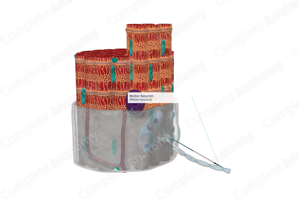
Motor neurons are usually made up of large and myelinated alpha-efferent axons. They arise from motor neuron cell bodies in the anterior horns of the gray matter within the spinal cord. Their terminal branches are short unmyelinated twigs which travel through the endomysium to form part of the neuromuscular junction.
Key Features/Anatomical Relations
The motor neuron axon branches into many terminal branches, which enter the endomysium. The myelin sheath of the axon ends at the neuromuscular junction, however, the terminal branches are covered by a thin layer, the neurilemma, which arises from the outer membrane of the Schwann cell. This external lamina of the Schwann cell, fuses with the sarcolemma, covering the points of contact between axonal branches and the muscle cell (Martini, Nath and Bartholomew, 2017).
The terminal axonal branches are dilated into a presynaptic structure which sits in shallow depressions on the surface of the muscle fiber. These areas are the receptor regions. The sarcolemma at these points will have deep junctional folds for increased surface area, ready to receive neurotransmitters responsible for muscle contraction.
Function
The function of the neuromuscular junction is to induce an action potential across the muscle fiber, initiating muscle contraction.
The nerve action potential travels across the axon of the motor neuron to its terminal branches and to the dilated presynaptic structures which sit within shallow depressions on the muscle fiber. These dilated ends contain both mitochondria and vesicles, the latter of which contain the neurotransmitter, acetylcholine. The nerve action potential triggers the release of acetylcholine which diffuses across the space between the presynaptic dilation and the sarcolemma. This space is called the synaptic cleft. The sarcolemma at these junctions contains a series of folds to increase the surface area as they contain acetylcholine receptors. Once the acetylcholine binds with their respective receptors, it induces an action potential across the muscle. The action potential will move along the surface of the muscle cell, along the t tubules that penetrate the sarcoplasm initiating release of CA2+ which in turn induces the contraction cycle (Standring, 2016).
List of Clinical Correlates
—Botulism
References
Dorland, W. (2011) Dorland's Illustrated Medical Dictionary. 32nd edn. Philadelphia, USA: Elsevier Saunders.
Martini, F. H., Nath, J. L. and Bartholomew, E. F. (2017) Fundamentals of Anatomy & Physiology. Pearson Education.
Standring, S. (2016) Gray's Anatomy: The Anatomical Basis of Clinical Practice. Gray's Anatomy Series: Elsevier Limited.
