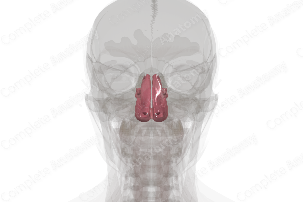
Structure
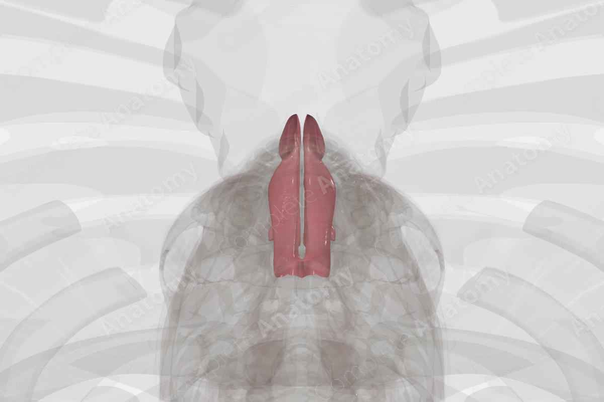
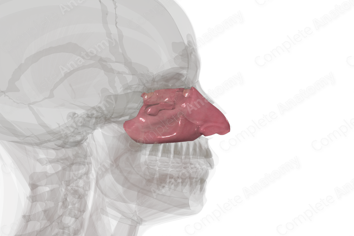
The nasal cavity is the passage from the external surface of the face, through the piriform aperture, to the nasopharynx, through the choanae. It has four walls composed mostly of bone and some cartilage. All walls are covered with mucosa lines with pseudostratified columnar epithelium (respiratory epithelium), except for a small, superior patch of olfactory epithelium containing olfactory receptors.
Related parts of the anatomy
Key Features & Anatomical Relations
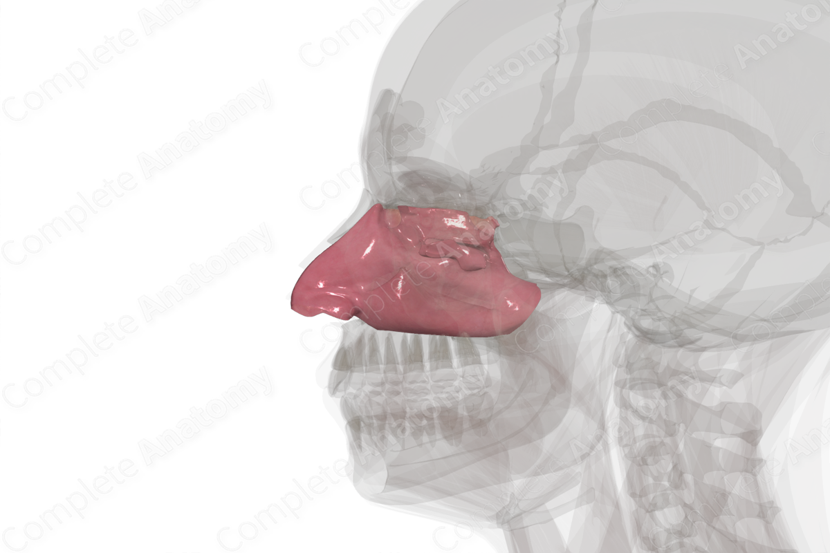
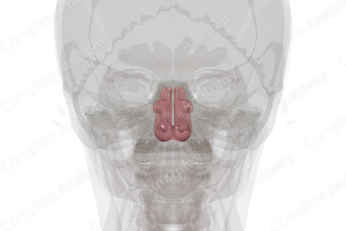
The medial wall of the nasal cavity is formed by the nasal septum. Anteriorly, it is continuous with the septal cartilage of the external nose. Posteriorly, it Is formed by the bony perpendicular plate of ethmoid (superiorly) and the vomer (inferiorly). The cribriform plate of the ethmoid bone forms much of the narrow roof of the nasal cavity, with small contributions from nasal, frontal, and sphenoid bones. The floor of the cavity is formed by the horizontal plate of the ethmoid bone, anteriorly, and the palatine process of the maxilla, posteriorly.
The lateral wall of the nasal cavity is formed of several bones, roughly, from anterior to posterior: frontal process of maxilla, lacrimal, ethmoid, perpendicular plate of ethmoid, and medial pterygoid plate of sphenoid. This wall contains many significant features. Three “shelves” of bone, convex inferiorly, extend medially. These form the superior, middle, and inferior nasal conchae (turbinates). The inferior concha is the final bony component of the lateral wall of the nasal cavity, being a bone in its own right. It articulates with the ethmoid bone. Inferior and tucked under the conchae are spaces called meatuses. Several passages open into each meatus. Specifically, the inferior meatus receives drainage from the lacrimal sac, and the middle and superior meatuses from paranasal sinuses.
List of Clinical Correlates
- Sinusitis
Learn more about this topic from other Elsevier products

Nasal cavity and larynx histology: Video, Anatomy

Master Nasal cavity and larynx histology: anatomy, function, and structure. High-yield review with labeled diagrams, videos, and practice quizzes.
