Quick Facts
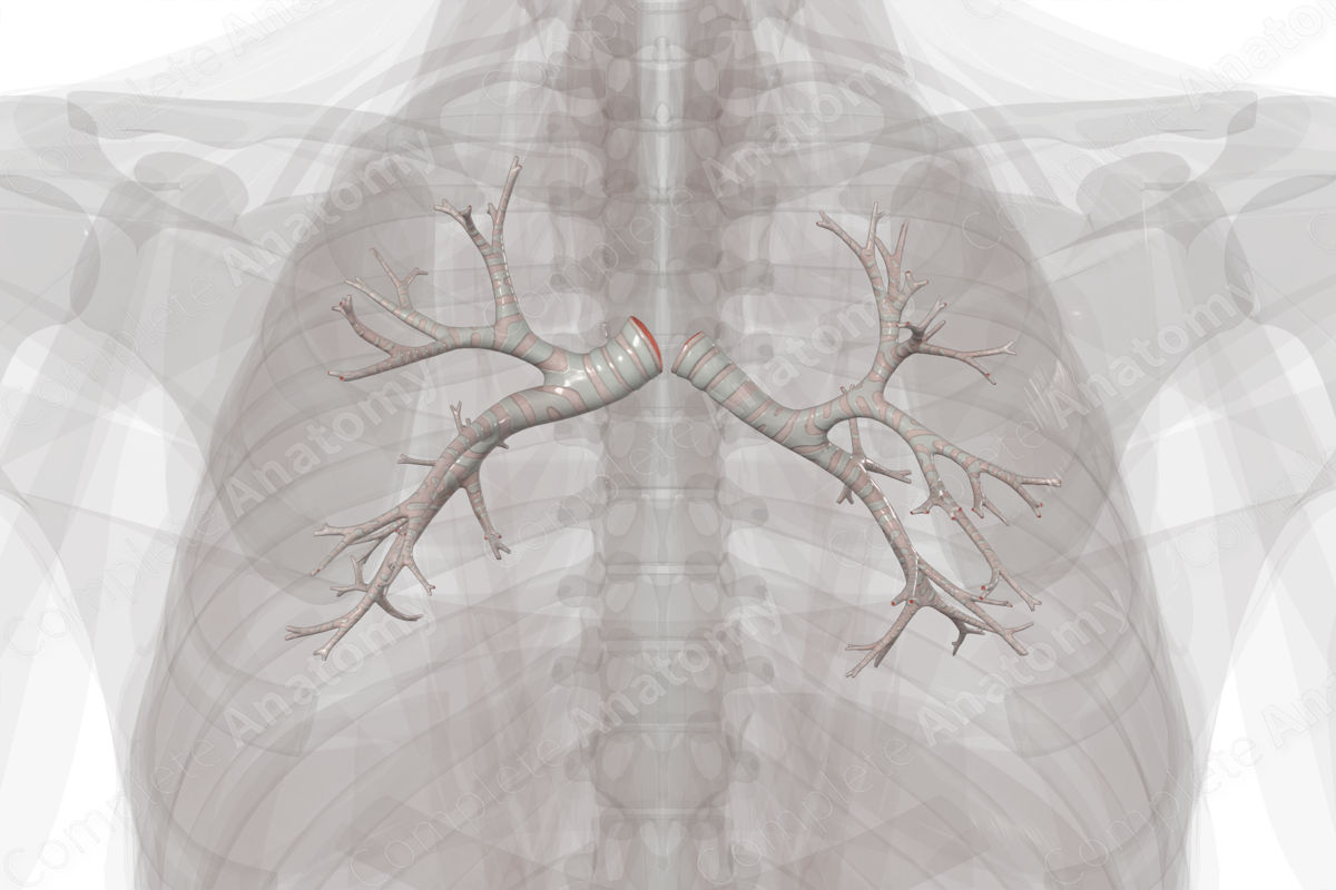
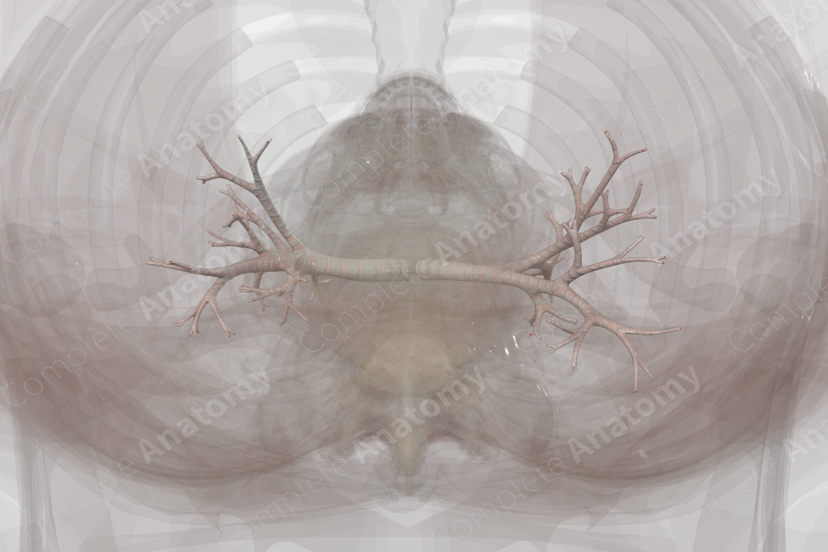
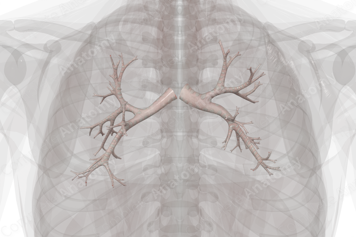
Location: Begin at the bifurcation of the trachea at the sternal angle (T4/T5) and enters the hilum of each lung.
Arterial Supply: Bronchial arteries.
Venous Drainage: Bronchial and pulmonary veins.
Innervation: Anterior and posterior pulmonary plexuses.
Lymphatic Drainage: Intrapulmonary lymph nodes.
Related parts of the anatomy
Structure
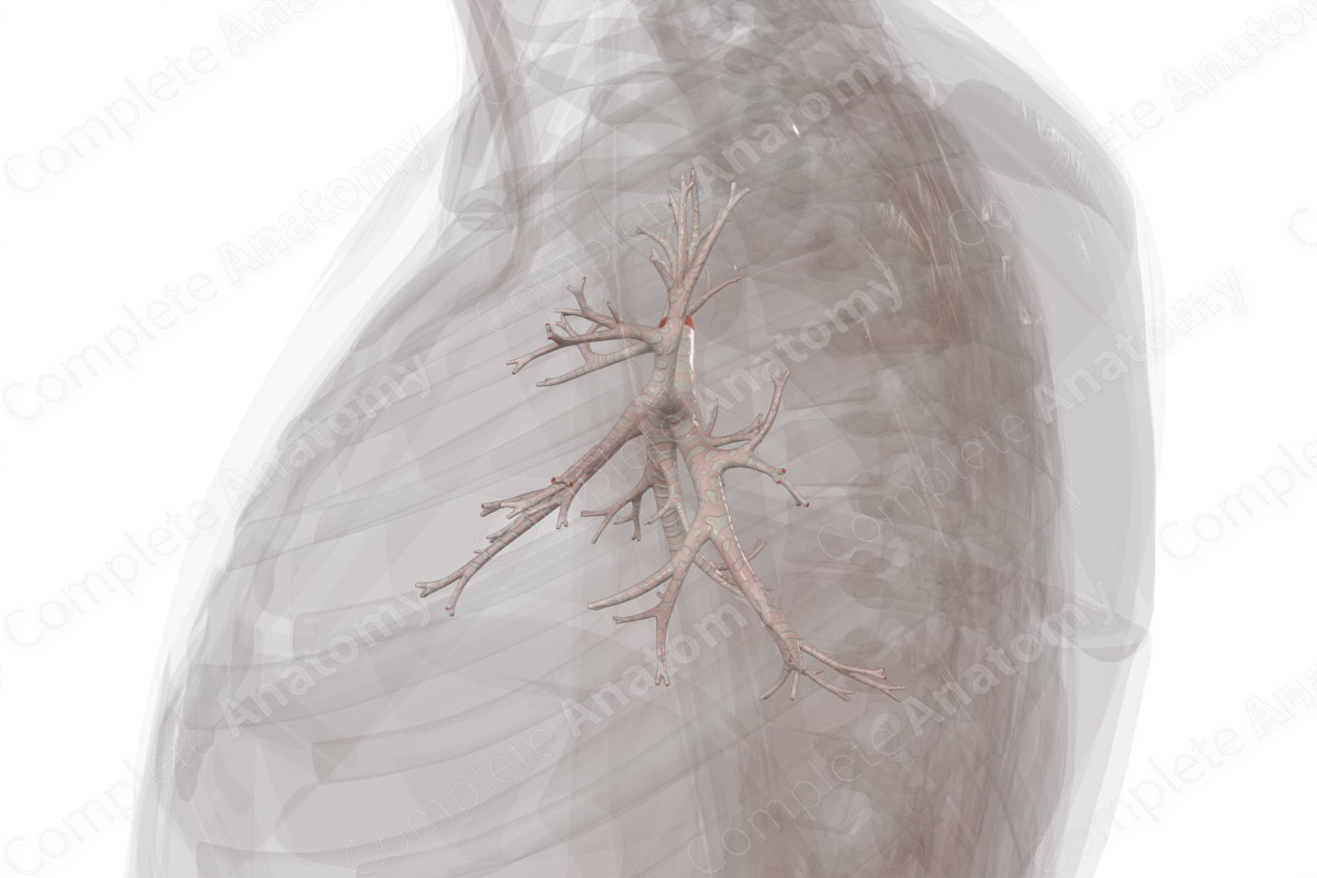
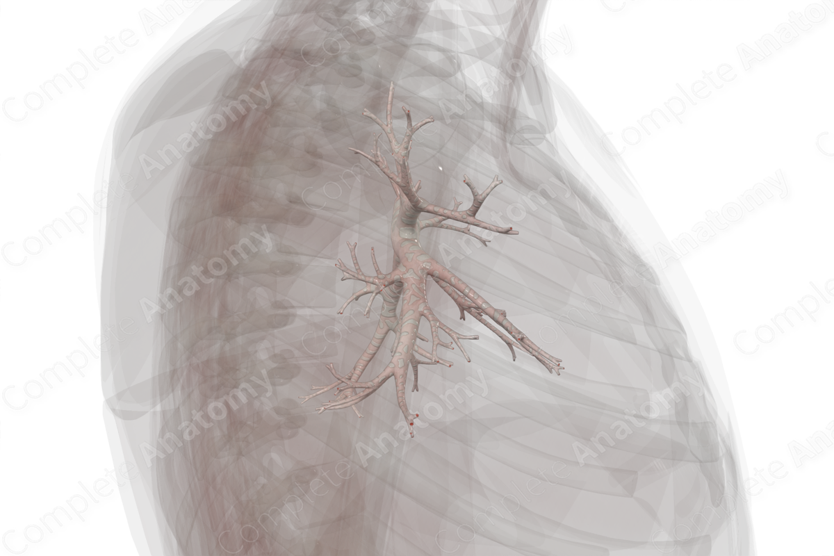
The bronchi arise from the bifurcation of the trachea into the right and left main bronchi at the level of the sternal angle (or transverse thoracic plane or angle of Louis). The main bronchi bifurcate as they enter the hila of each lung, forming lobar bronchi (or secondary bronchi) that enter the individual lobes of each lung. Within the lobes, the lobar bronchi continue to divide into segmental (or tertiary) bronchi that supply the bronchopulmonary segments of each lobe.
The segmental bronchi continue to divide, producing 20–25 generations of conducting bronchioles that eventually end as the terminal bronchioles and have an internal diameter of 0.5-1mm. Bronchioles also undergo extensive branching, with their distal-most branches ending in alveolar ducts and alveoli.
Like the trachea, the walls of the bronchi consist of an inner mucosal layer, with an underlying submucosal layer. Surrounding the submucosal layer is an irregular configuration of rings of hyaline cartilage, along with smooth muscle fibers. Towards the terminal parts of the segmental bronchi and their transition into terminal bronchioles, the cartilaginous rings are replaced by hyaline cartilage plates. The walls of the bronchioles have no hyaline cartilage, and their patency is determined by the tone of the smooth muscle within its walls (Eroschenko, 2008).
The bronchi can be divided into two portions: the conducting part and the respiratory part. The conducting part includes the primary, secondary and tertiary bronchi and the terminal bronchioles, along with the trachea, and is responsible for conducting air to and from the lungs. The respiratory part includes the respiratory bronchioles, alveolar ducts, alveolar sacs and alveoli proper. The respiratory portion may conduct air but is also responsible for gaseous exchange. The respiratory portion of the bronchi can be seen in our micro-anatomy model of the bronchial tree.
Anatomical Relations
The main, lobar, and segmental (or primary, secondary, and tertiary) bronchi are accompanied by a pulmonary artery, which delivers deoxygenated blood to the lungs, and a pulmonary vein, which drains newly oxygenated blood from the lungs. Therefore, each lobe and each bronchopulmonary segment of the lung has its own bronchus, pulmonary artery, and adjacent intersegmental vein. This arrangement is important during surgery, since a single segment can be resected without affecting an adjacent segment.
Function
The primary function of the bronchi is to conduct air to the lungs so that gas exchange and oxygenation of blood can occur. As the bronchi are constantly exposed to inhaled allergens, particles, and microbes, it is necessary to clear these threats to maintain homeostasis. The mucosa of the bronchi and bronchioles play the vital role in protecting the lungs from these environmental insults, thus, maintaining homeostasis (Ganesan, Comstock and Sajjan, 2013).
Arterial Supply
The bronchial arteries supply the oxygenated blood and nutrients to the bronchi. A paired group of left bronchial branches (superior and inferior bronchial branches) arise directly from the thoracic aorta. A right bronchial branch arises indirectly from the thoracic aorta via the right-sided posterior intercostal artery, usually the third right posterior intercostal artery.
Venous Drainage
The bronchial veins receive blood from the bronchi, hilar structures, such as lymph nodes, and visceral pleura adjacent to the hilum. A left and right bronchial vein arise from the posterior surface of the bronchi. These veins travel posteriorly to their terminations at the azygos vein on the right and the hemiazygos vein, or sometimes the superior intercostal vein, on the left. Additionally, some blood from the bronchial arteries is drained by the pulmonary veins. Please see our micro-anatomy model of the tracheobronchial tree for more details.
Innervation
The bronchi receive autonomic innervation from the anterior and posterior pulmonary plexuses. The nerves of the pulmonary plexuses originate from the thoracic branches of the upper sympathetic trunk (sympathetic contribution) and the vagus nerve (parasympathetic contribution). For both sympathetic and parasympathetic fibers, axons may travel directly to the pulmonary plexus, or may travel through the cardiac plexus first. The autonomic innervation of the trachea controls airway function, such as tone of smooth muscle, mucus secretion, and vascular permeability and blood flow.
Lymphatic Drainage
The intrapulmonary lymph nodes are located along the bronchi. These nodes drain the deep bronchopulmonary plexus located within the bronchopulmonary segments of the lungs. The intrapulmonary lymph nodes send efferents to the bronchopulmonary lymph nodes at the hilum of the lungs.
References
Eroschenko, V. P. (2008) DiFiore's Atlas of Histology with Functional Correlations. Point (Lippincott Williams and Wilkins) Series: Wolters Kluwer Health/Lippincott Williams & Wilkins.Ganesan, S., Comstock, A. T. and Sajjan, U. S. (2013) 'Barrier function of airway tract epithelium', Tissue Barriers., 1(4), pp. e24997.
Learn more about this topic from other Elsevier products
Trachea and bronchi histology: Video and Anatomy

Master Trachea and bronchi histology: anatomy, function, and structure. High-yield review with labeled diagrams, videos, and practice quizzes.
