Quick Facts
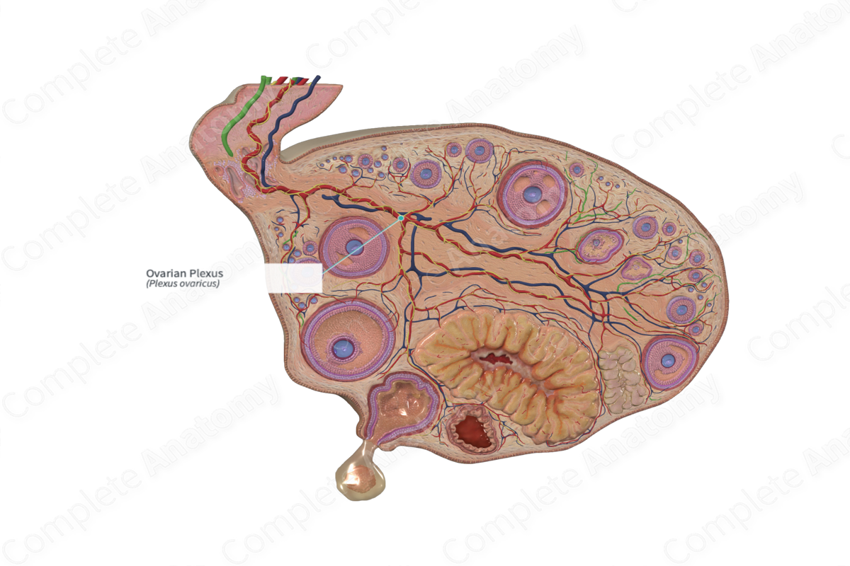
Sympathetic Contribution: Greater and lesser thoracic splanchnic (T10–T11) nerves, traveling through the abdominal and prevertebral autonomic ganglia and plexuses.
Parasympathetic Contribution: Pelvic splanchnic nerves (S2-S4).
Course: Follows the course of the ovarian artery to reach the pelvis.
Sympathetic Supply: Ovary, distal uterine tube, and uterine fundus.
Parasympathetic Supply: Ovary, distal uterine tube, and uterine fundus.
Related parts of the anatomy
Contributing Nerves
Sympathetic nerves descend with the ovarian artery in a fold of peritoneum that forms the suspensory ligament of the ovary. Some fibers pass through the superior and inferior hypogastric plexuses. Parasympathetic fibers cross the pelvic floor to the inferior hypogastric and ovarian plexuses.
Course
Descending sympathetic fibers work their way down the posterior abdominal wall in aortic, renal, periarterial, and ureteric plexuses. Those from the latter two plexuses follow the ovarian artery within the suspensory ligament of the ovary. Ascending sacral and pelvic splanchnic nerves pass along the pelvic floor. Some fibers ascend within the broad ligament with the uterine artery. Others continue medially to the inferior and superior hypogastric plexuses.
Branches
There are no named branches.
Supplied Structures
The ovarian plexus is thought to act as a vasoconstrictor (sympathetic) or as a vasodilator (parasympathetic) to the blood vessels within the ovary. Autonomic innervation is not required for ovulation and the fibers do not reach the ovarian follicles.
In addition, some sensory fibers travel within the ovarian plexus and convey pain from the ovaries to the central nervous system. This established nerve supply accounts for the high sensitivity of the ovaries to pressure and may present in the form of periumbilical pain (Oats and Abraham, 2017).
References
Oats, J. J. N. and Abraham, S. (2017) Llewellyn-Jones Fundamentals of Obstetrics and Gynaecology E-Book. 10th edn.: Elsevier Health Sciences.
