Structure and/or Key Feature(s)
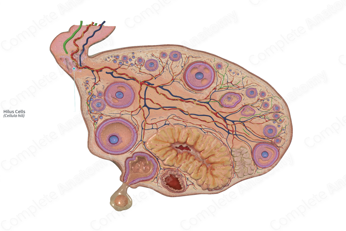
Ovarian hilus cells are located in the hilum of the ovary. Like the interstitial (Leydig) cells of the male gonads, hilar cells in the ovary contain Reinke crystalloids, lipofuscin pigment associated with protein production. They associated with vascular spaces and nonmyelinated nerve fibers.
Related parts of the anatomy
Function
Hilus cells may secrete androgens. They respond to hormonal changes during pregnancy and the onset of menopause.
List of Clinical Correlates
Tumors associated with hilus cells usually lead to masculinization (Ross and Pawlina, 2006).
References
Ross, M. H. and Pawlina, W. (2006) Histology: A text and atlas. Lippincott Williams & Wilkins
