Quick Facts
Origin: Pons.
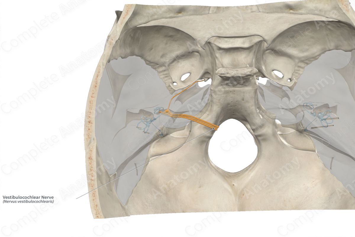
Course: Runs laterally from the pontomedullary junction through the internal acoustic meatus and into the inner ear.
Branches: Vestibular and cochlear nerves.
Supply: Special sensory innervation, carrying sensory information for hearing and balance.
Related parts of the anatomy
Origin
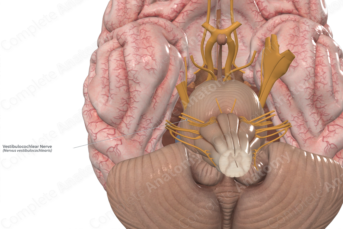
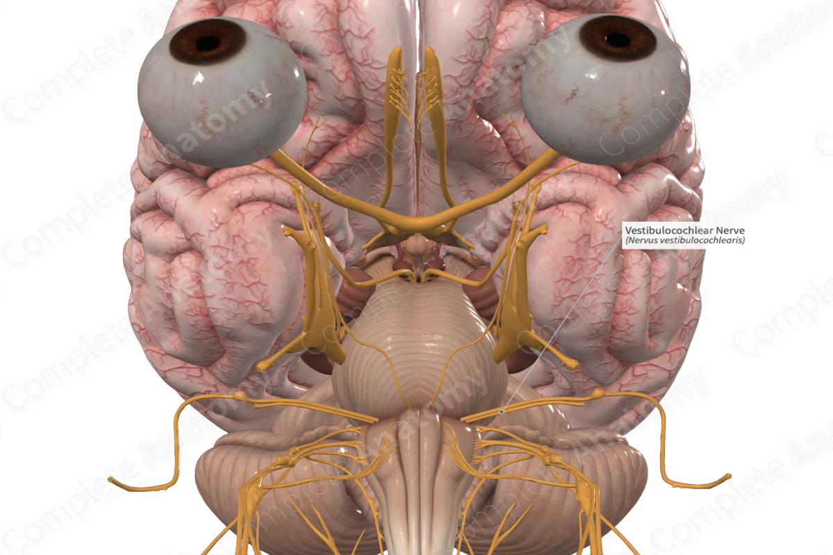
The vestibulocochlear nerve fibers originate from vestibular and cochlear nuclei which are found in the pons and medulla. The fibers emerge from the brainstem at the mediolateral pontomedullary junction, just lateral to the nervus intermedius and facial nerve.
Course
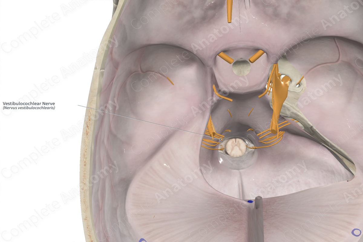
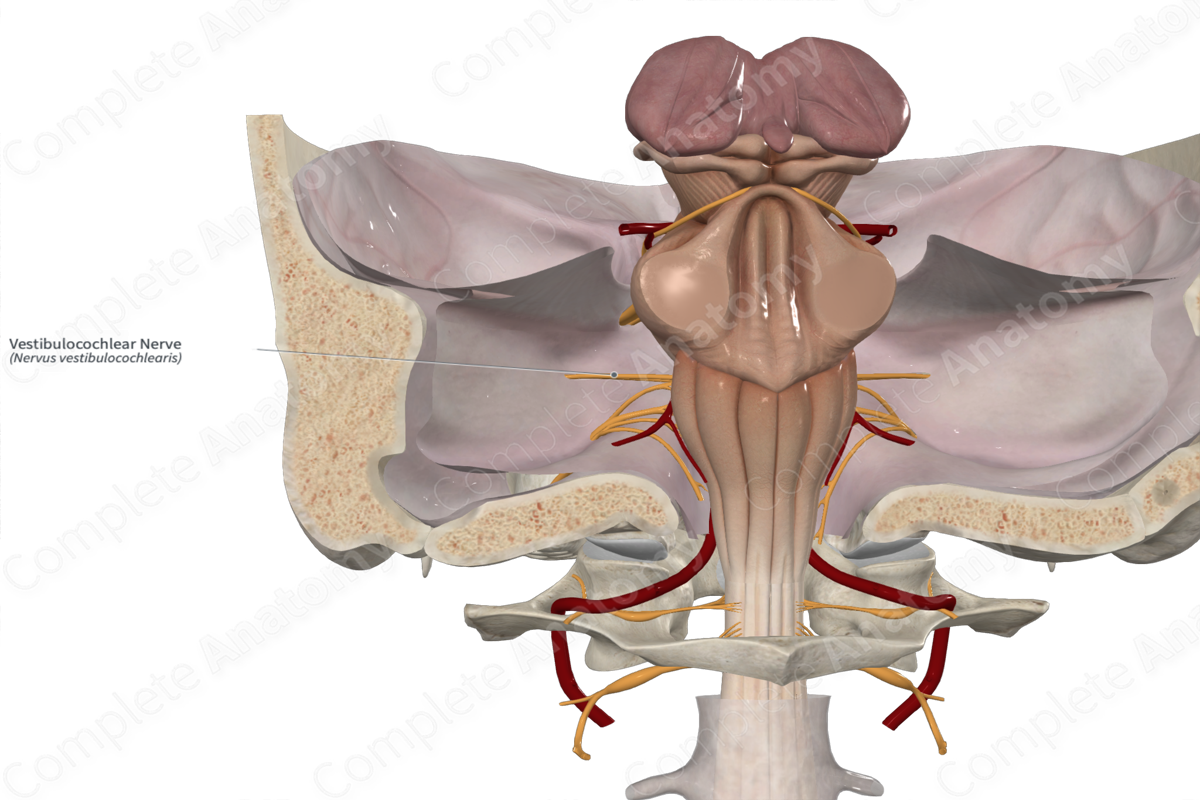
The vestibulocochlear nerve runs from the pontomedullary junction laterally past the cerebellopontine angle to the internal acoustic meatus, along with both the facial nerve and the nervus intermedius.
From the internal acoustic meatus, the branches of the vestibulocochlear nerve travel to the cochlea or the vestibular apparatus within the inner ear.
Branches
The vestibulocochlear nerve splits to give rise to two major nerves, the vestibular nerve and the cochlear nerve.
Supplied Structures
The vestibulocochlear nerve is a sensory nerve. Afferent fibers from the cochlea convey auditory information to the brainstem. Afferent fibers originating in the vestibular apparatus, the semicircular canals and statoconium (otolith), convey sensory information regarding head movements to the brainstem.
List of Clinical Correlates
—Vertigo
—Nystagmus
—Disequilibrium
—Deafness
Learn more about this topic from other Elsevier products

Anatomy of the vestibulocochlear nerve (CN VIII)

Master Anatomy of the vestibulocochlear nerve (CN VIII): structure, function, and composition. High-yield review.
