Quick Facts
Location: Trigeminal (Mekel’s cave).
Branches: The trigeminal ganglion is the site at which the ophthalmic, maxillary, and mandibular nerves merge to form the trigeminal nerve proper.
Supply: Sensory innervation from the forehead and face, paranasal sinuses, nasal and oral cavities, and anterior two thirds of the tongue.
Related parts of the anatomy
Location
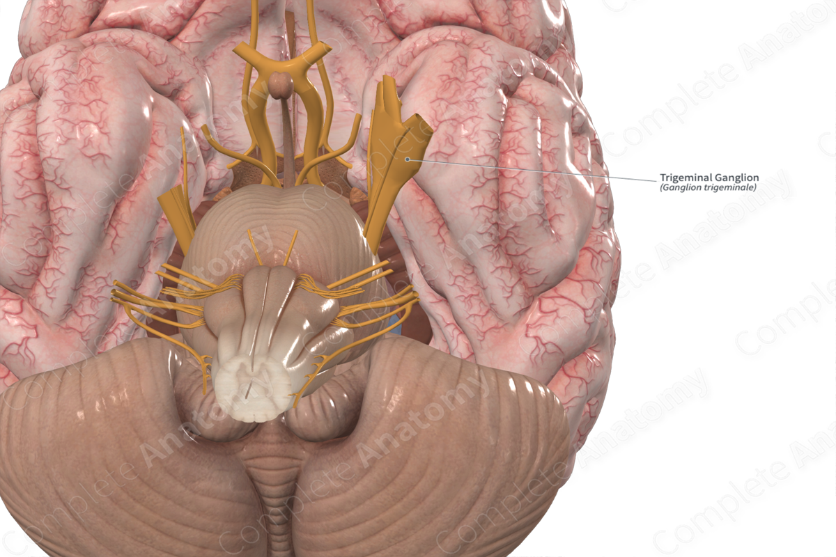
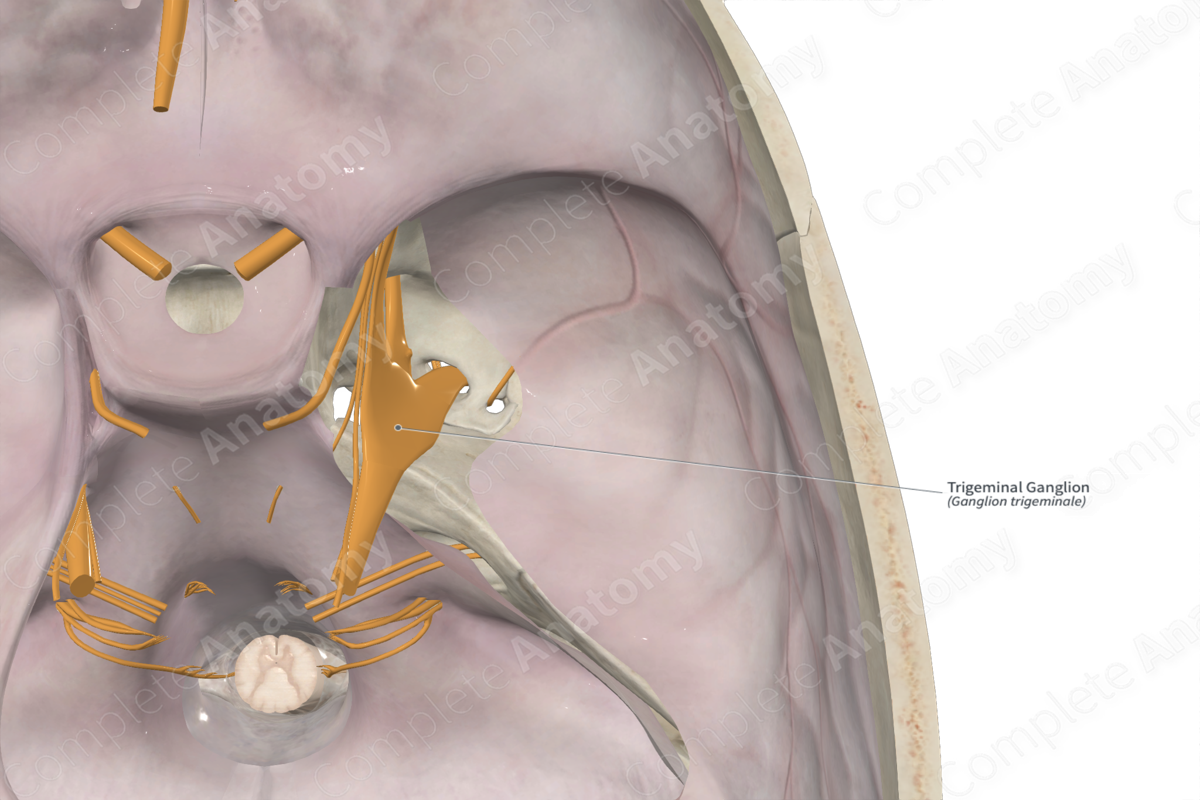
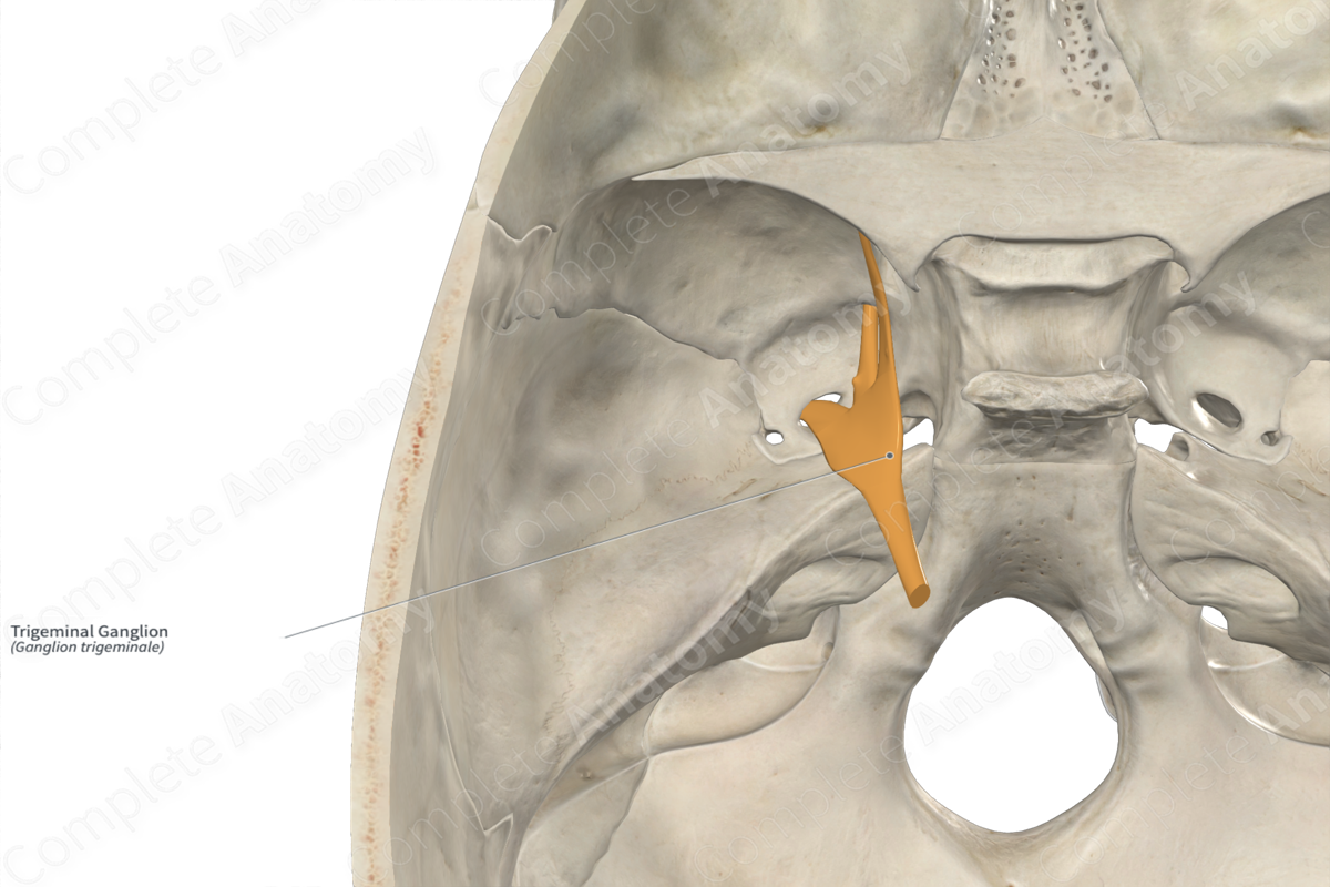
The trigeminal ganglion sits in a slight depression on the lateral side of the petrous ridge of the temporal bone, just posterior to the anterior most part of the ridge. It is located just posterior to the foramen lacerum where the carotid canal ends. This depression is called the trigeminal impression and because the trigeminal ganglion is enveloped in dura, the depression is typically not very large.
The trigeminal ganglion is enveloped between the periosteal and meningeal layers of the dura mater, and its anterior half extends into the posterior part of the cavernous sinus. These dural layers involute back on themselves to form a cavity called the trigeminal (or Mekel’s) cave.
The trigeminal ganglion is a neural crest derived ganglion that contains the sensory neurons of the trigeminal nerve. It can be thought of as equivalent to the dorsal root ganglia of the spinal nerves (O'Rahilly & Müller, 2007).
Branches
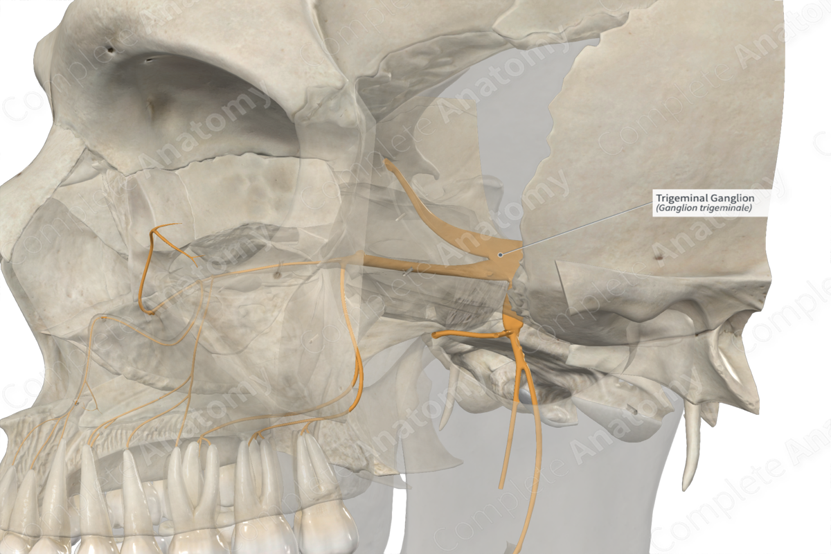
Medially, the trigeminal ganglion is the site of the trigeminal nerve. This consists of the large sensory root of the trigeminal nerve and a much smaller motor root of the trigeminal nerve which is found just ventral to the sensory root.
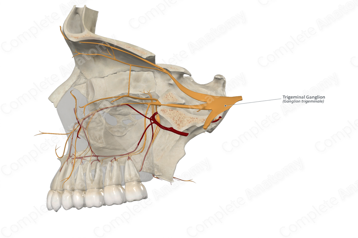
Anteriorly and laterally, the trigeminal ganglion gives rise to three branches, the ophthalmic, maxillary, and mandibular nerves.
Supplied Structures
The trigeminal ganglion is a sensory structure that is the location of all sensory neurons of the trigeminal nerve. Its neurons convey sensation from the three branches of the trigeminal nerve:
The ophthalmic nerve travels into the orbit and conveys general sensory information from the skin of the upper face, roughly from the anterior nose and upper half of the eye, superiorly to the top of the head. This nerve also conveys sensation to the cornea, the mucosa of ethmoid air cells, frontal sinus, and sphenoid sinus, and small portions of the dural folds.
The maxillary nerve travels into the pterygopalatine fossa and innervates the skin of the middle face, roughly from the lower half of the eyes down to the upper lip with its territory arcing up as it moves posteriorly. Other tissues supplied by the maxillary nerve are the maxillary teeth and gingiva, the palate, the nasal cavity, the maxillary sinus, the nasopharynx, the lower lateral nose, and portions of the cranial dura mater.
The mandibular nerve travels into the infratemporal fossa and innervates the skin of the lower face, from the lower lips down to the lower margins of the mandible. Its territory arcs posteriorly and upward, just anterior to the ear. Other tissues innervated by sensory fibers of the mandibular nerve include the mucous membrane lining the cheek, mandibular gingiva, mandibular teeth, and anterior two thirds of the tongue.
List of Clinical Correlates
—Corneal reflex
—Trigeminal neuralgia
—Herpes zoster
References
O'Rahilly, R. & Müller, F. (2007) The development of the neural crest in the human. Journal of anatomy, 211(3), 335-351.
Learn more about this topic from other Elsevier products

