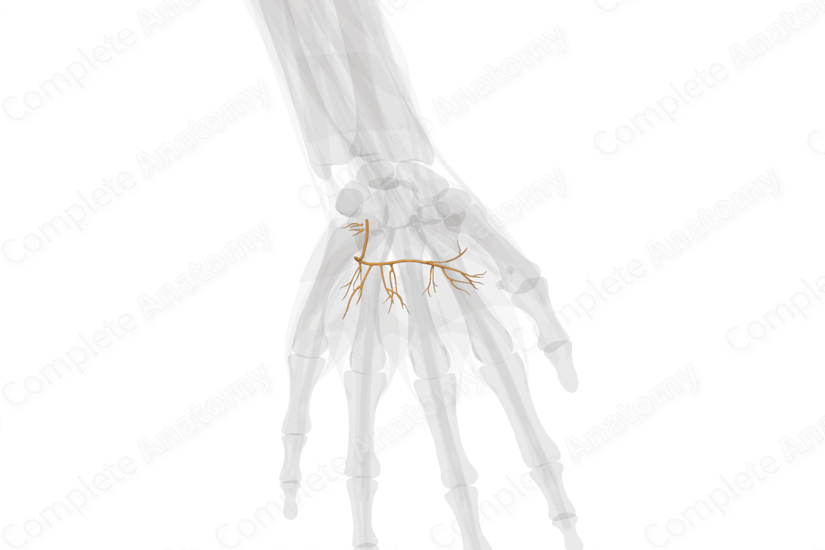
Description
The ulnar nerve crosses the wrist between the tendons of flexor carpi ulnaris and flexor digitorum superficialis muscles and divides into superficial and deep terminal branches at the distal border of the flexor retinaculum. Most of the intrinsic muscles of the hand are innervated by the deep branch of ulnar nerve (Polatsch et al., 2007).
The deep terminal branch of the ulnar nerve accompanies the deep branch of the ulnar artery. It passes backwards between the abductor and flexor digiti minimi, and then between the opponens digiti minimi and the fifth metacarpal bone, lying on the hook of the hamate. Finally, it turns laterally within the concavity of the deep palmar arch and supplies the medial two lumbricals and eight interosseous muscles, as the nerve crosses the palm. The deep branch innervates most of the intrinsic muscles of the hand. It terminates by supplying the adductor pollicis, and occasionally the deep head of flexor pollicis brevis.
The superficial branch of the ulnar nerve innervates only the palmaris brevis muscle. The remainder provides cutaneous innervation.
References
Polatsch, D. B., Melone, C. P., Beldner, S. and Incorvaia, A. (2007) 'Ulnar nerve anatomy', Hand Clin, 23(3), pp. 283-9, v.
Learn more about this topic from other Elsevier products


