Quick Facts
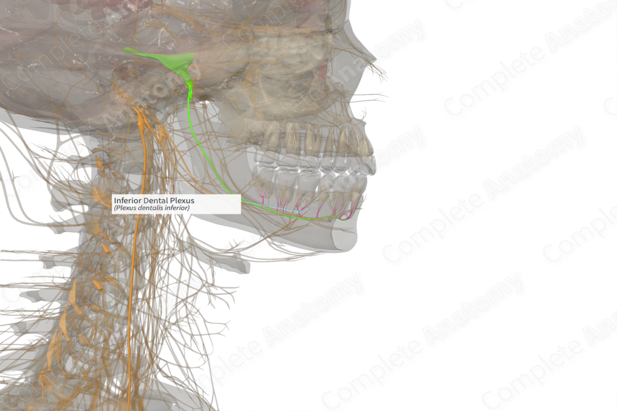
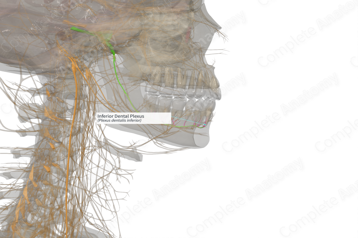
Origin: Inferior alveolar nerve.
Course: Runs superiorly through the alveolar portion of the body of the mandible to the roots of the molar and premolar mandibular teeth.
Branches: Inferior dental and gingival branches.
Supply: Conveys general sense fibers from the molar and premolar mandibular teeth, as well as related periodontal membranes and buccal gingiva.
Related parts of the anatomy
Origin
The inferior dental plexus originates as branches of the inferior alveolar nerve. They branch off the inferior alveolar nerve as it passes down the mandibular canal past the molar and premolar teeth of the mandible.
Its sensory fibers have cell bodies located in the trigeminal ganglion.
Course
The inferior dental plexus ascends through the alveolar bone of the mandible to reach the roots of the mandibular molars and premolars, as well as surrounding tissues.
Branches
The inferior dental plexus gives rise to inferior dental and gingival branches.
Supplied Structures
The inferior dental plexus conveys general sense fibers from the mandibular molars and premolars, as well as the periodontal tissue and buccal gingiva associated with these teeth.
Learn more about this topic from other Elsevier products

