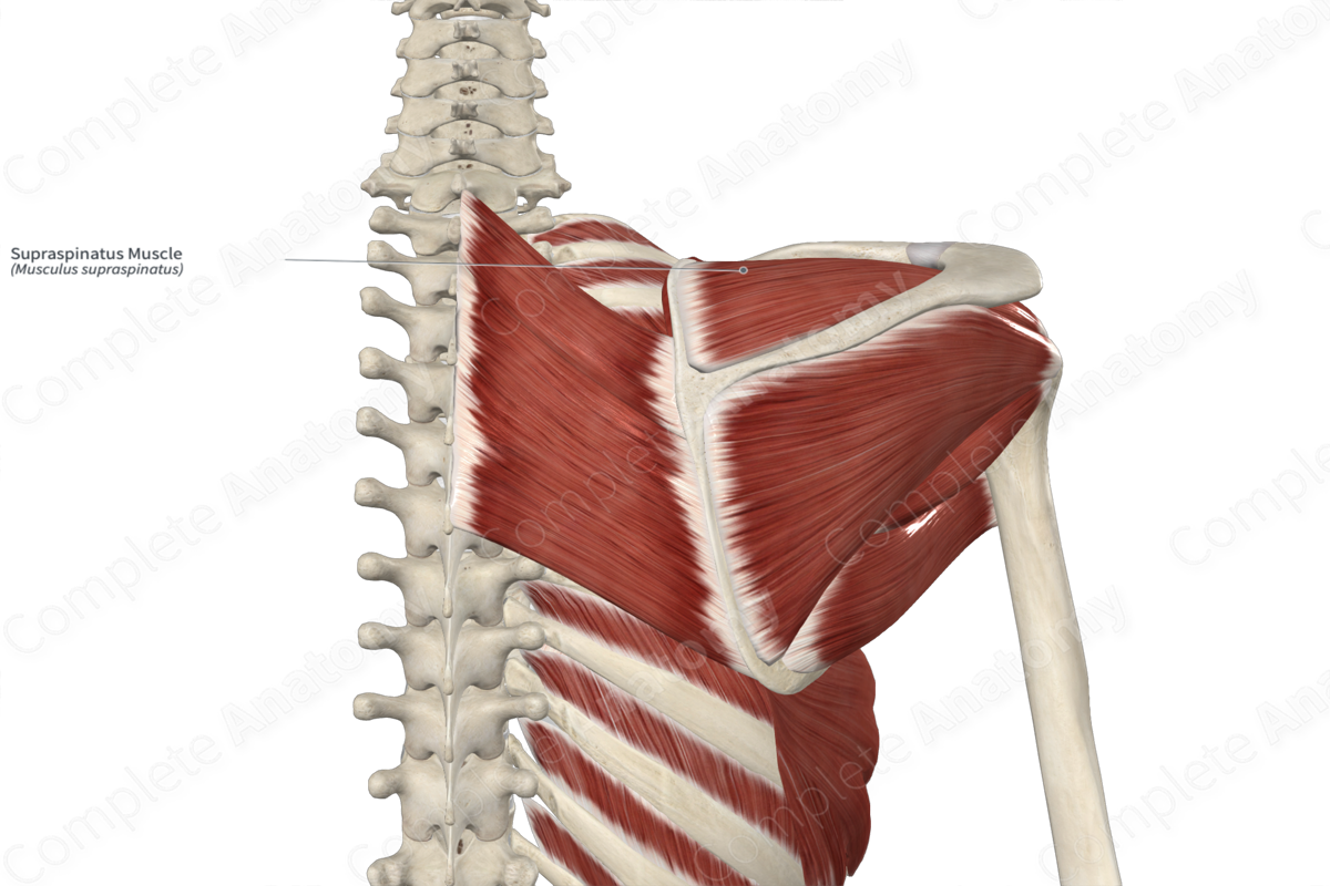
Quick Facts
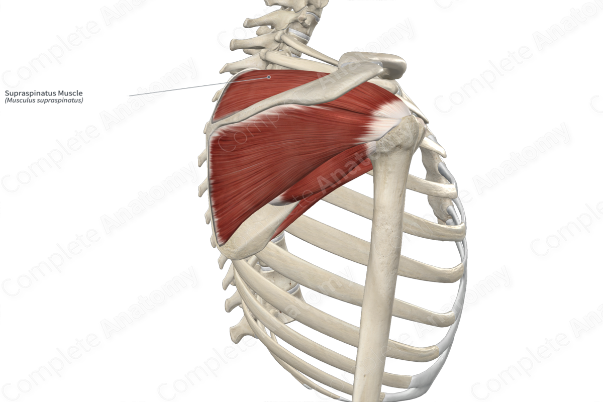
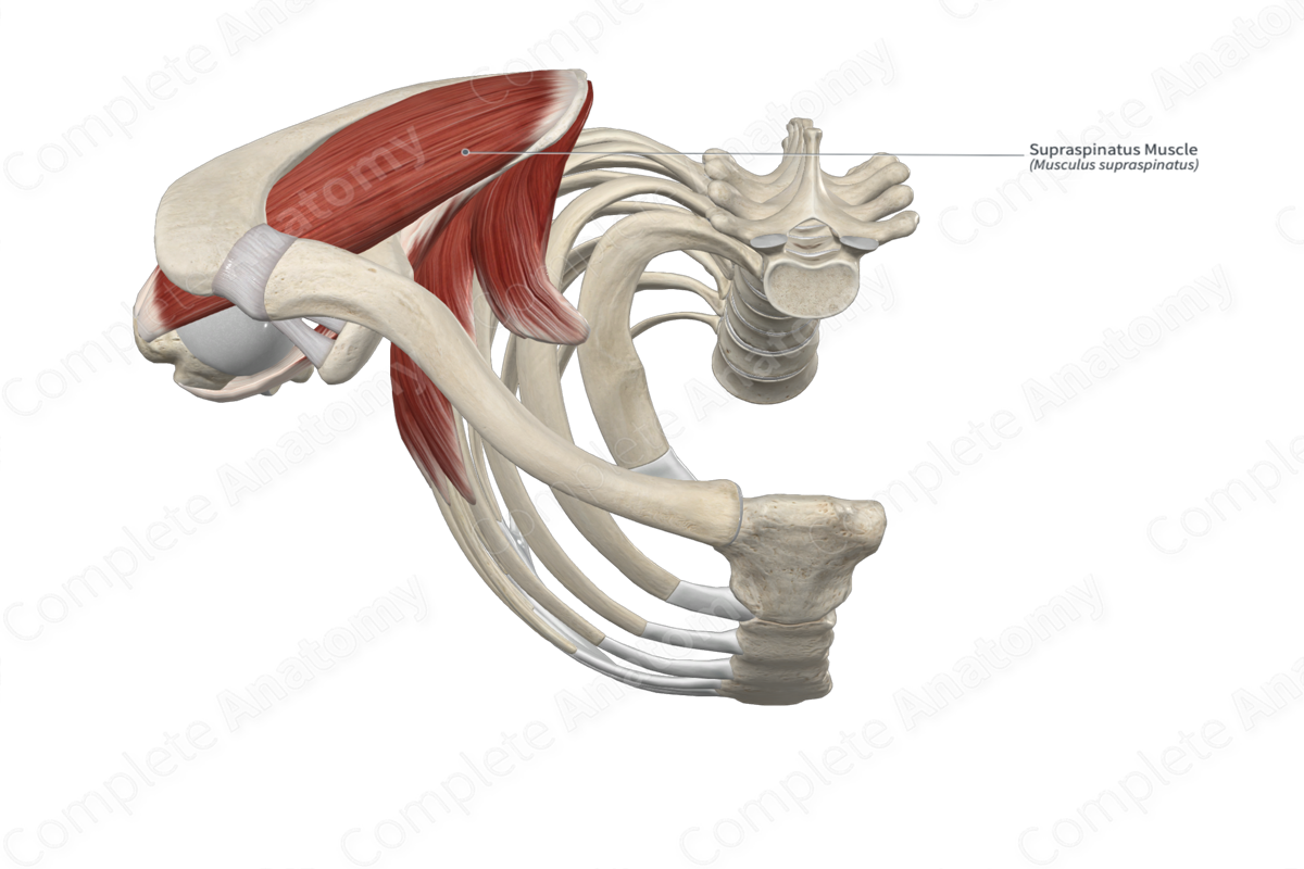
Origin: Supraspinous fossa of scapula.
Insertion: Greater tubercle of humerus.
Action: Abducts and stabilizes arm at glenohumeral (shoulder) joint.
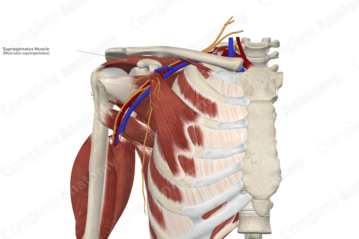
Innervation: Suprascapular nerve (C5-C6).
Arterial Supply: Suprascapular and dorsal scapular arteries.
Related parts of the anatomy
Origin
The supraspinatus muscle originates from the:
medial two thirds of the supraspinous fossa of the scapula;
internal surface of the supraspinous fascia.
Insertion
The fibers of the supraspinatus muscle travel laterally and insert, via a thick tendon, onto the superior facet of the greater tubercle of the humerus. Part of this tendon also merges with the capsule of the glenohumeral (shoulder) joint.
Key Features & Anatomical Relations
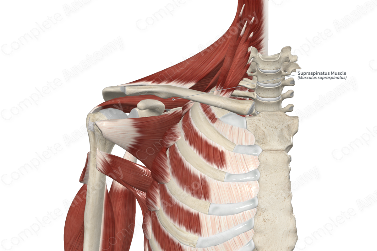
The supraspinatus muscle is one of the rotator cuff muscles. It is a thick, bipennate type of skeletal muscle. It is located:
anterior (deep) to the trapezius muscle and the supraspinous fascia;
posterior (superficial) to the scapula, and the subtendinous bursa of supraspinatus muscle.
Actions & Testing
The supraspinatus muscle abducts the arm at the glenohumeral (shoulder) joint. It is one of the four rotator cuff (SITS) muscles, the other three being the infraspinatus, teres minor and subscapularis muscles. These muscles work together to stabilize the glenohumeral joint, by holding the head of the humerus in the glenoid fossa of the scapula, during its movements (Standring, 2016).
The supraspinatus muscle can be tested by abducting the arm at the glenohumeral joint against resistance, starting from the fully adducted position, during which the muscle can be palpated (Sinnatamby, 2011).
List of Clinical Correlates
Injury or rupture of rotator cuff
References
Sinnatamby, C. S. (2011) Last's Anatomy: Regional and Applied. ClinicalKey 2012: Churchill Livingstone/Elsevier.
Standring, S. (2016) Gray's Anatomy: The Anatomical Basis of Clinical Practice. Gray's Anatomy Series 41st edn.: Elsevier Limited.
Learn more about this topic from other Elsevier products

