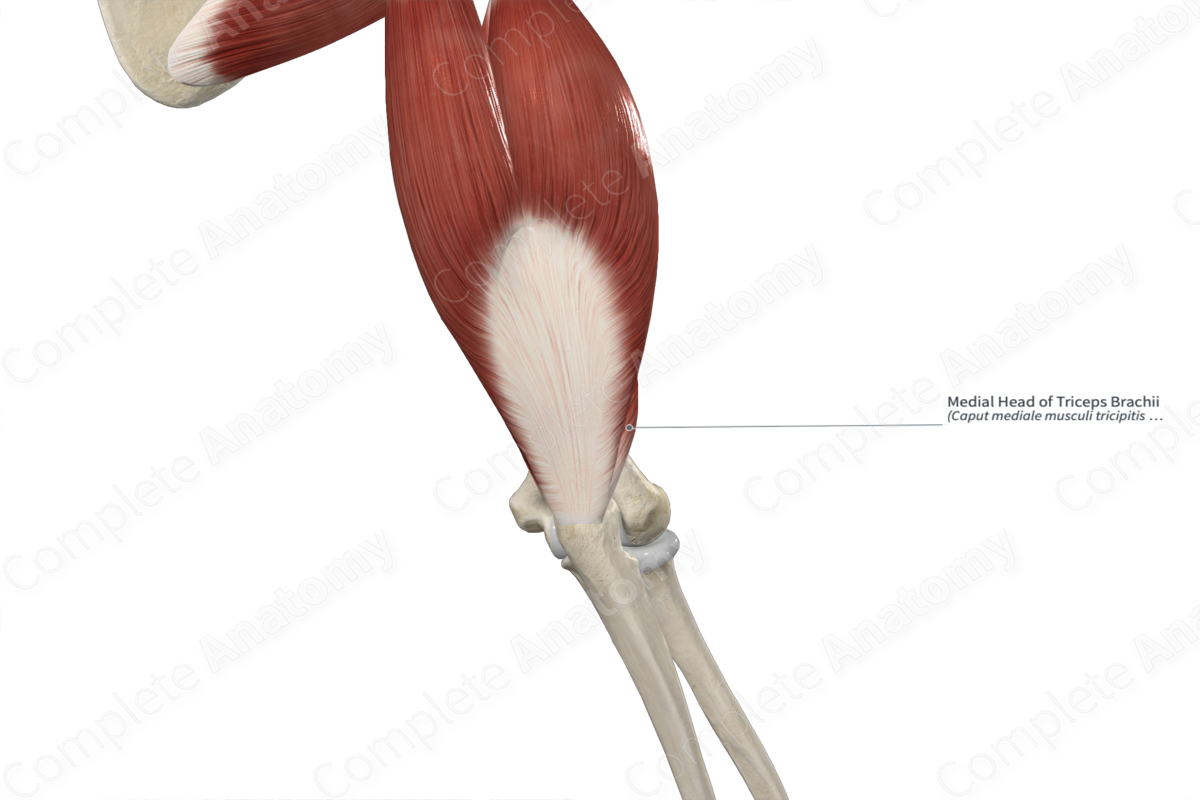
Quick Facts
Origin: Posterior surface of humerus, inferior to radial groove.
Insertion: Olecranon of ulna and adjacent antebrachial fascia.
Action: Extends forearm at elbow joint.
Innervation: Radial nerve (C8).
Arterial Supply: Deep brachial and superior ulnar collateral arteries.
Related parts of the anatomy
Origin
The medial head of triceps brachii muscle originates from an extensive area on the posterior surface of the humerus that is located inferior to the groove for radial nerve. It also originates from the medial and lateral intermuscular septa of the arm.
Insertion
The fibers of the medial, lateral, and long heads of triceps brachii muscle all converge to a single triceps brachii tendon, which inserts onto both the superior end of the olecranon of ulna and the antebrachial fascia. Some fibers from the medial head of the triceps brachii muscle attach to the articular capsule of the elbow joint. These fibers, which by some, are considered an additional muscle in the arm known as the articularis cubiti muscle (or subanconeus).
Key Features & Anatomical Relations
The triceps brachii muscle is found in the posterior compartment of the arm. It is a fusiform type of skeletal muscle and is composed of three heads: medial, lateral, and long, where:
- the medial head is located deep to the lateral and long heads;
- the long head is located medial to the lateral head.
The triceps brachii muscle is located:
- posterior (superficial) to the deep brachial artery and radial nerve;
- lateral to the teres major and teres minor muscles.
Actions & Testing
The triceps brachii muscle extends the forearm at the elbow joint. This action is not affected by pronation or supination of the forearm. The medial head is active in the presence or absence of resistance.
The triceps brachii muscle can be tested by extending the forearm at the elbow joint against resistance, during which it can be palpated (Standring, 2016).
References
Standring, S. (2016) Gray's Anatomy: The Anatomical Basis of Clinical Practice. Gray's Anatomy Series 41st edn.: Elsevier Limited.
Learn more about this topic from other Elsevier products


