
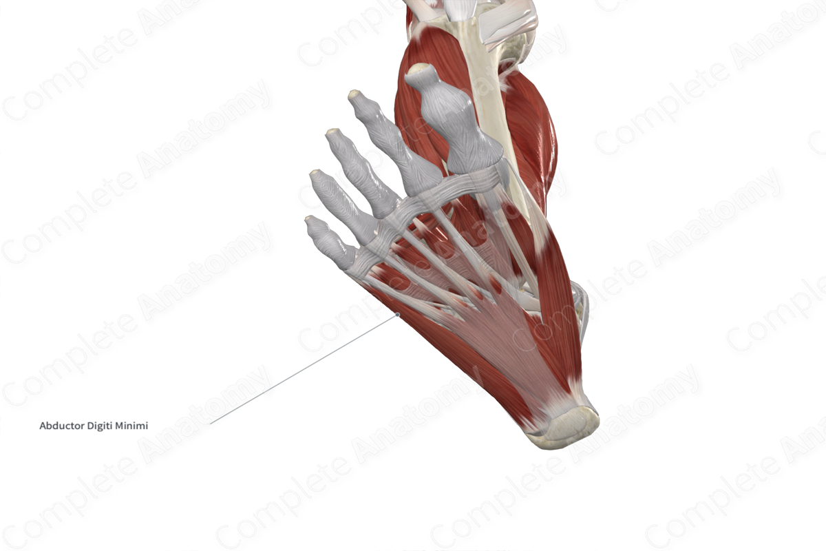
Quick Facts
Origin: Medial and lateral processes of calcaneal tuberosity and plantar aponeurosis.
Insertion: Lateral aspect of base of proximal phalanx of little toe.
Action: Abducts and flexes little toe at its metatarsophalangeal joint.
Innervation: Lateral plantar nerve (S2-S3).
Arterial Supply: Lateral plantar, proper plantar digital, and plantar metatarsal arteries.


Related parts of the anatomy
Origin
The abductor digiti minimi muscle of foot originates from the:
- medial and lateral processes of the calcaneal tuberosity;
- plantar aponeurosis;
- adjacent intermuscular septum.
Insertion
The fibers of the abductor digiti minimi muscle of foot travel anterolaterally and insert onto the lateral aspect of the base of the proximal phalanx of little toe.
Key Features & Anatomical Relations
The abductor digiti minimi muscle of foot is located in the first layer of muscles that are found in the plantar part of the foot. It is a short, fusiform type of skeletal muscle that contributes to the formation of the lateral margin of the foot.
It is located:
- superficial (inferior) to the calcaneus and fifth metatarsal bone, the quadratus plantae muscle, and the flexor digiti minimi muscle of foot;
- lateral to the plantar aponeurosis, flexor digitorum brevis muscle, flexor digiti minimi muscle of foot, lateral plantar vessels, and lateral plantar nerve.
Actions
The abductor digiti minimi muscle of foot is involved in multiple actions:
- abducts the proximal phalanx of little toe (i.e., draws it away from the longitudinal axial line of the second toe) at its metatarsophalangeal joint (Sinnatamby, 2011);
- flexes the proximal phalanx of little toe at its metatarsophalangeal joint (Standring, 2016).
References
Sinnatamby, C. S. (2011) Last's Anatomy: Regional and Applied. ClinicalKey 2012: Churchill Livingstone/Elsevier.
Standring, S. (2016) Gray's Anatomy: The Anatomical Basis of Clinical Practice. Gray's Anatomy Series 41st edn.: Elsevier Limited.
Learn more about this topic from other Elsevier products

