Quick Facts
Origin: Ischial tuberosity, linea aspera, and lateral supracondylar line.
Insertion: Head of fibula.
Action: Flexes and laterally rotates leg at knee joint; extends thigh at hip joint.
Innervation: Tibial (L5-S2) and common fibular (L5-S2) divisions of sciatic nerve.
Arterial Supply: Perforating arteries of deep femoral artery, inferior gluteal and medial circumflex femoral arteries.
Related parts of the anatomy
Origin
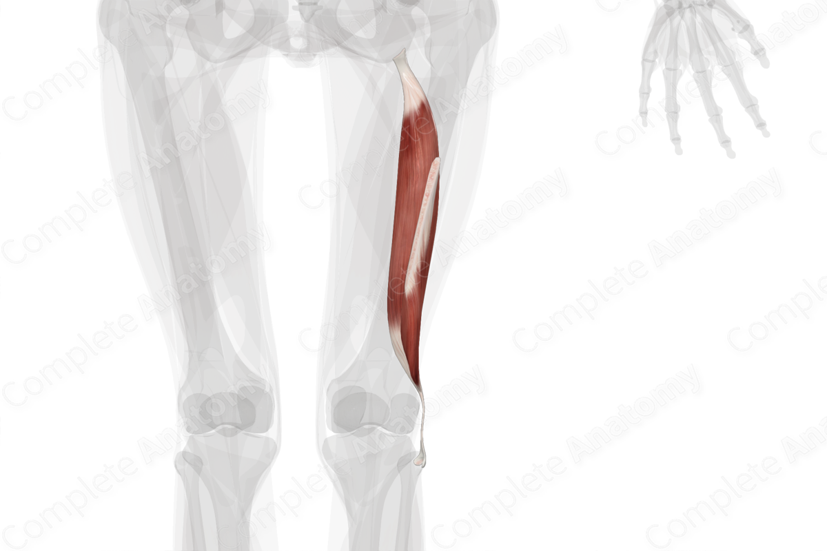
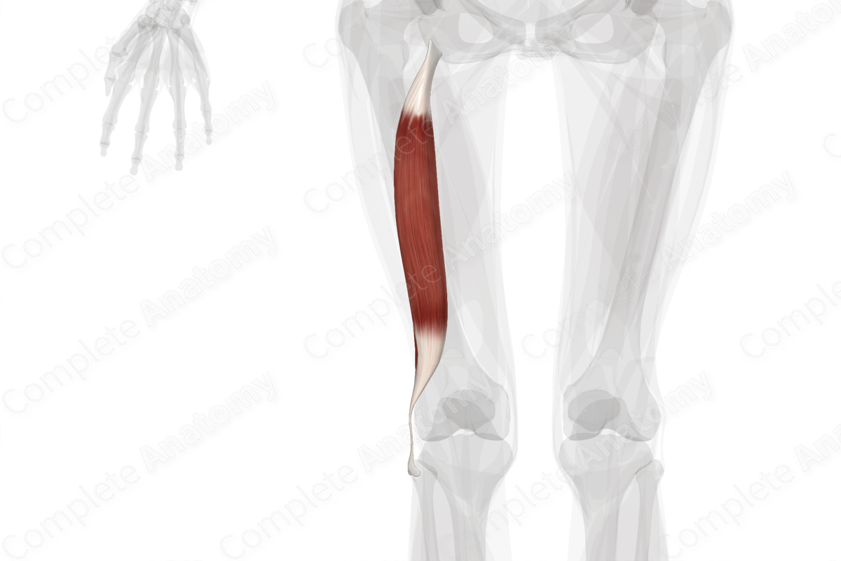
The biceps femoris muscle consists of two heads:
- a long head, which originates from the medial aspect of the posterior portion of the ischial tuberosity;
- a short head, which originates from the lateral lip of linea aspera and lateral supracondylar line of the femur.
Insertion
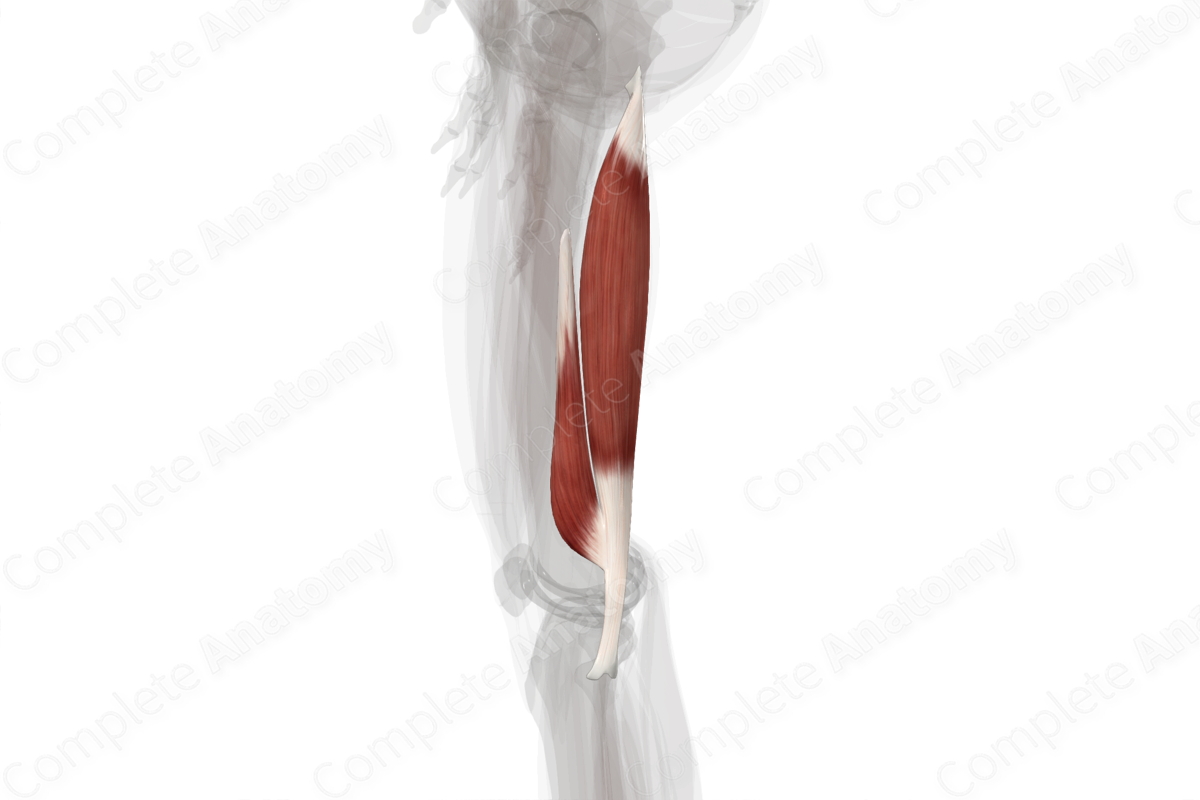
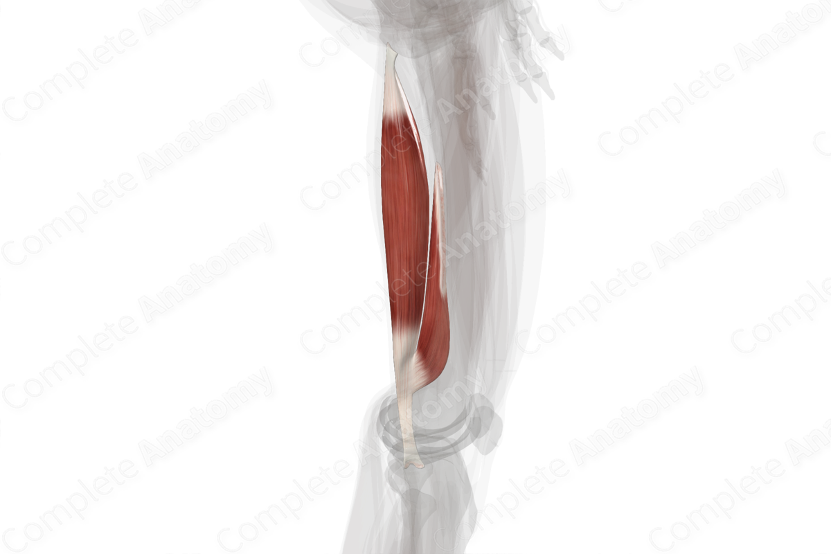
The fibers of the long and short heads of biceps femoris travel inferiorly and converge to a single biceps femoris tendon. This tendon splits around the fibular collateral ligament and then inserts onto the lateral aspect of the head of the fibula.
Key Features & Anatomical Relations
Overall, the biceps femoris muscle is found in the posterior compartment of the thigh. It is a fusiform type of skeletal muscle and is composed of a medially located long head and a laterally located short head.
The biceps femoris muscle is located:
- anterior to the gluteus maximus muscle (at its proximal end);
- posterior to the femur, adductor magnus and vastus lateralis muscles, and the sciatic nerve;
- medial to the iliotibial tract;
- lateral to the semitendinosus and semimembranosus muscles.
Overall, the biceps femoris muscle contributes to the formation of the popliteal fossa, where the muscle and tendon form its superolateral boundary. The term “hamstrings” is the collective name given to the long head of biceps femoris, semitendinosus, and semimembranosus muscles. These three muscles share similar features, including:
- originating from the ischial tuberosity;
- acting on both the hip and knee joints;
- innervated by the tibial division of sciatic nerve.
Actions & Testing
Overall, the biceps femoris is involved in multiple actions:
- flexes the leg at the knee joint;
- laterally rotates the leg at the knee joint while this joint is held in a semiflexed position;
- extends the thigh at the hip joint.
The biceps femoris muscle cannot be tested in isolation, therefore all of the muscles of the posterior compartment of the thigh are tested simultaneously by flexing the leg at the knee joint against resistance while lying in the prone position, during which the biceps femoris tendon can be seen and palpated (Standring, 2016).
List of Clinical Correlates
- Hamstring strain
- Hamstring tear
- Avulsion of ischial tuberosity
- Biceps femoris tendinitis
References
Standring, S. (2016) Gray's Anatomy: The Anatomical Basis of Clinical Practice. Gray's Anatomy Series 41st edn.: Elsevier Limited.
Learn more about this topic from other Elsevier products
Biceps Femoris: What Is It, Location, Action, and More

The biceps femoris is a long muscle in the posterior compartment of the thigh responsible for movement at both the hip and knee joints. Learn with Osmosis

