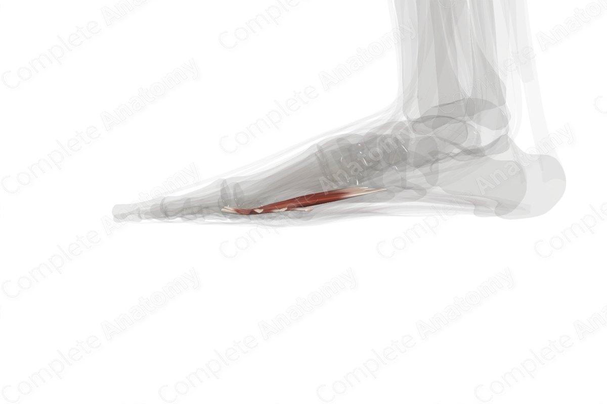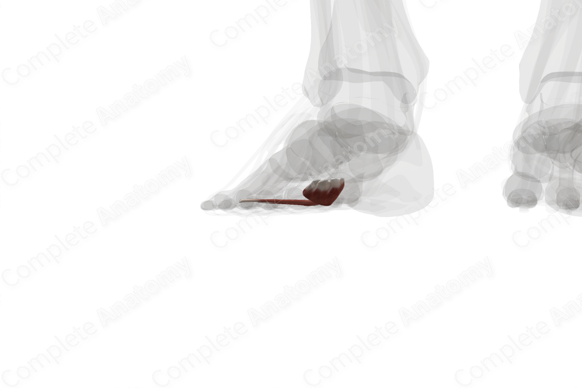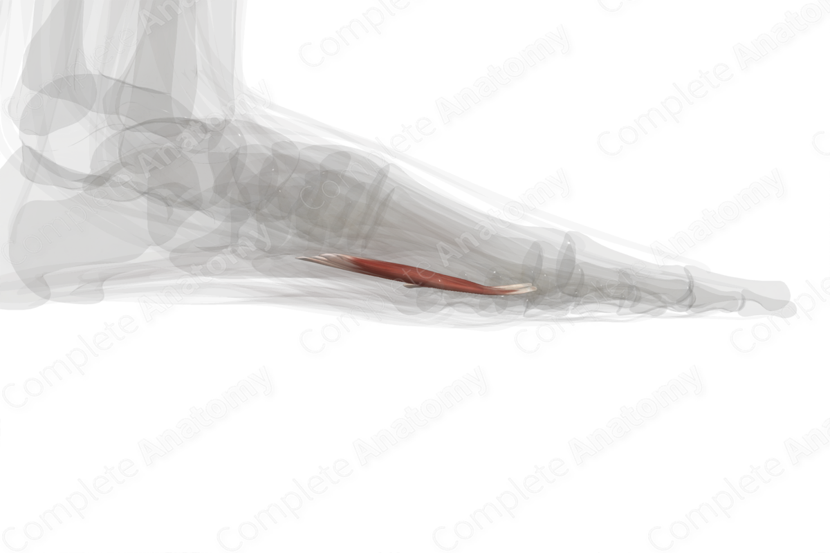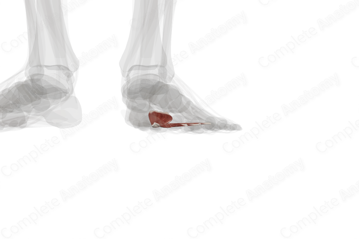
Quick Facts
Origin: Plantar aspects of bases of second to fourth metatarsal bones, tendon of fibularis longus muscle, plantar ligaments of metatarsophalangeal joints, and deep transverse metatarsal ligament.
Insertion: Lateral aspect of base of proximal phalanx of great toe.
Action: Adducts great toe at its metatarsophalangeal joint.
Innervation: Deep branch of lateral plantar nerve (S2-S3).
Arterial Supply: Medial and lateral plantar arteries, deep plantar arch, and plantar metatarsal arteries.
Related parts of the anatomy
Origin
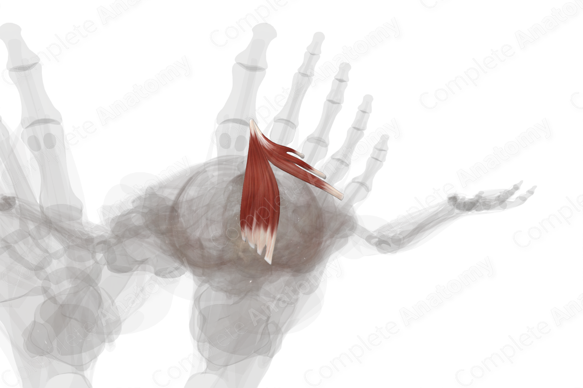
The adductor hallucis muscle consists of two heads:
- the oblique head, which originates from the plantar aspect of the bases of the second to fourth metatarsal bones and the distal end of the tendon of fibularis longus muscle;
- the transverse head, which originates from the plantar ligaments of the metatarsophalangeal joints of the third, fourth and little toes, and the adjacent deep transverse metatarsal ligament.
Insertion
The muscle bellies of the oblique and transverse heads of adductor hallucis travel anterolaterally and converge to a single tendon, which inserts onto the lateral aspect of the base of the proximal phalanx of the great toe.
Key Features & Anatomical Relations
Overall, the adductor hallucis muscle is located in the third layer of muscles that are found in the plantar part of the foot. It is a fan-shaped skeletal muscle and is composed of two heads, which are named based on the orientation of their muscle fibers:
- a large oblique head;
- a small transverse head.
The adductor hallucis muscle is located:
- superior to the first to third lumbrical muscles of foot and the tendons of the flexor digitorum longus and flexor digitorum brevis muscles;
- inferior to the second to fourth metatarsal bones, the first and second dorsal interossei and first and second plantar interossei muscles of the foot;
- lateral to the flexor hallucis brevis muscle.
Actions
Overall, the adductor hallucis muscle is involved in multiple actions:
- adducts the proximal phalanx of great toe (i.e., draws it towards the longitudinal axial line of the second toe) at the first metatarsophalangeal joint;
- helps stabilize the transverse arch of the foot (Sinnatamby, 2011).
List of Clinical Correlates
- Hallux valgus
References
Sinnatamby, C. S. (2011) Last's Anatomy: Regional and Applied. ClinicalKey 2012: Churchill Livingstone/Elsevier.
Learn more about this topic from other Elsevier products

