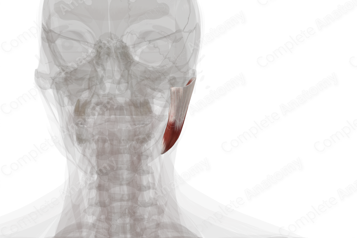
Quick Facts
Origin: Inferior aspect of zygomatic arch.
Insertion: Lateral aspect of coronoid process and ramus of mandible.
Action: Elevates mandible; assists in protraction of mandible.
Innervation: Masseteric nerve (CN V3).
Arterial Supply: Masseteric, transverse facial, and facial arteries.
Related parts of the anatomy
Origin
The masseter muscle arises from the inferior aspect of the zygomatic arch.
Insertion
The masseter muscle inserts into the lateral aspect of the coronoid process and ramus of the mandible.
Key Features & Anatomical Relations
The posterior aspect of the masseter muscle sits deep to the parotid gland. The masseter muscle is crossed anteriorly by the parotid duct, branches of the facial nerve, and the transverse facial artery. A buccal fat pad sits between the anterior margin of the masseter and the buccinator muscle.
Actions
Overall, the masseter muscle is involved in multiple actions:
- elevates the mandible at the temporomandibular joint;
- assists in protraction of the mandible at the temporomandibular joint (Standring, 2016).
List of Clinical Correlates
- Submasseteric space infections
- Trismus
References
Standring, S. (2016) Gray's Anatomy: The Anatomical Basis of Clinical Practice. Gray's Anatomy Series 41st edn.: Elsevier Limited.
Learn more about this topic from other Elsevier products


