Structure
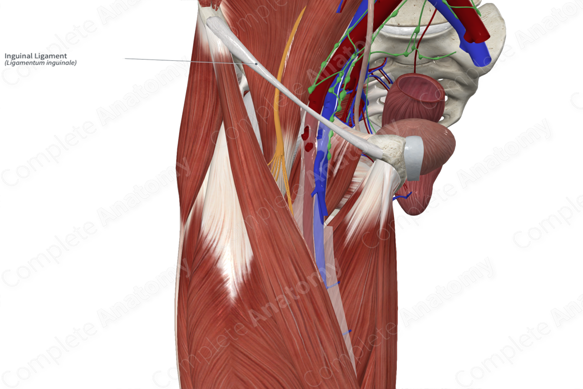
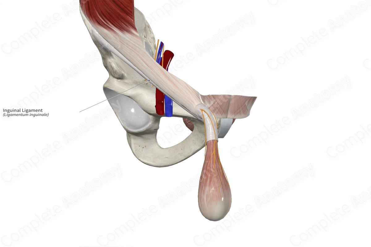
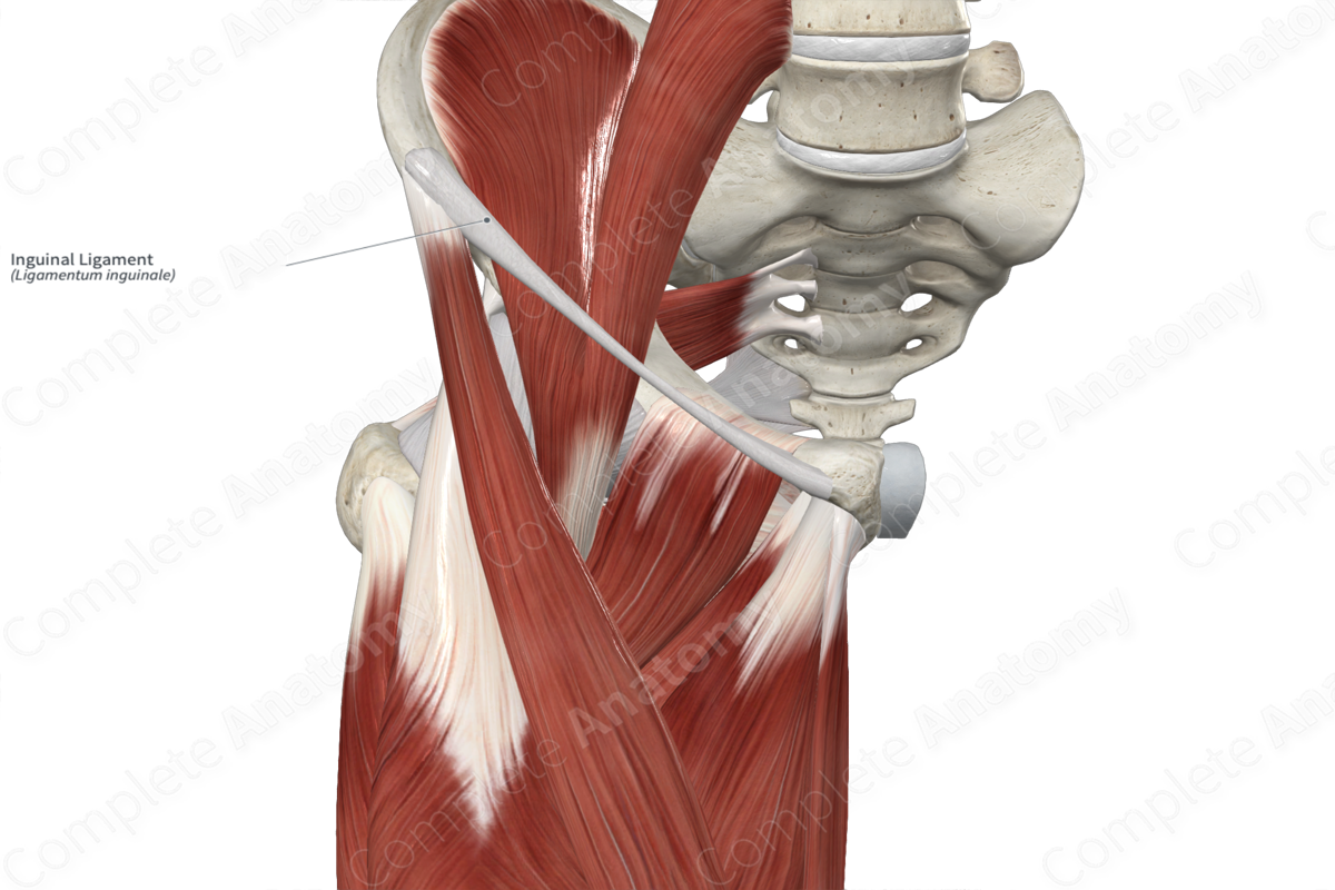
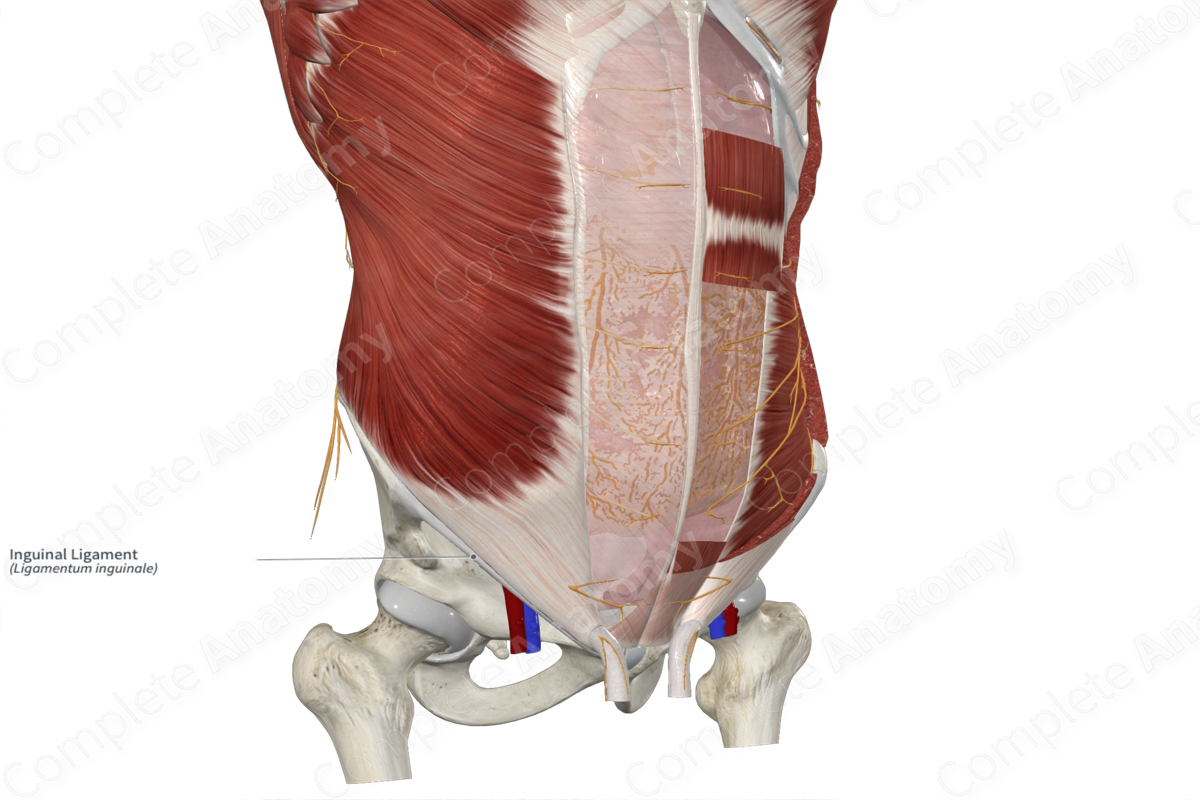
The inguinal ligament is the thickened, inferior border of the external abdominal oblique muscle aponeurosis. It has an inferior and anterior convexity, therefore is not exactly linear. It merges with the iliopsoas fascia laterally and the fascia lata of the thigh anteriorly. Medially, fibers from the inguinal ligament extend posteriorly and attach to the pectineal line. This forms a shelf-life triangular ligament, the lacunar ligament. Other reflected, medial fibers of the inguinal ligament run posterosuperiorly to the superficial ring to merge with the rectus sheath and linea alba.
Related parts of the anatomy
Anatomical Relations
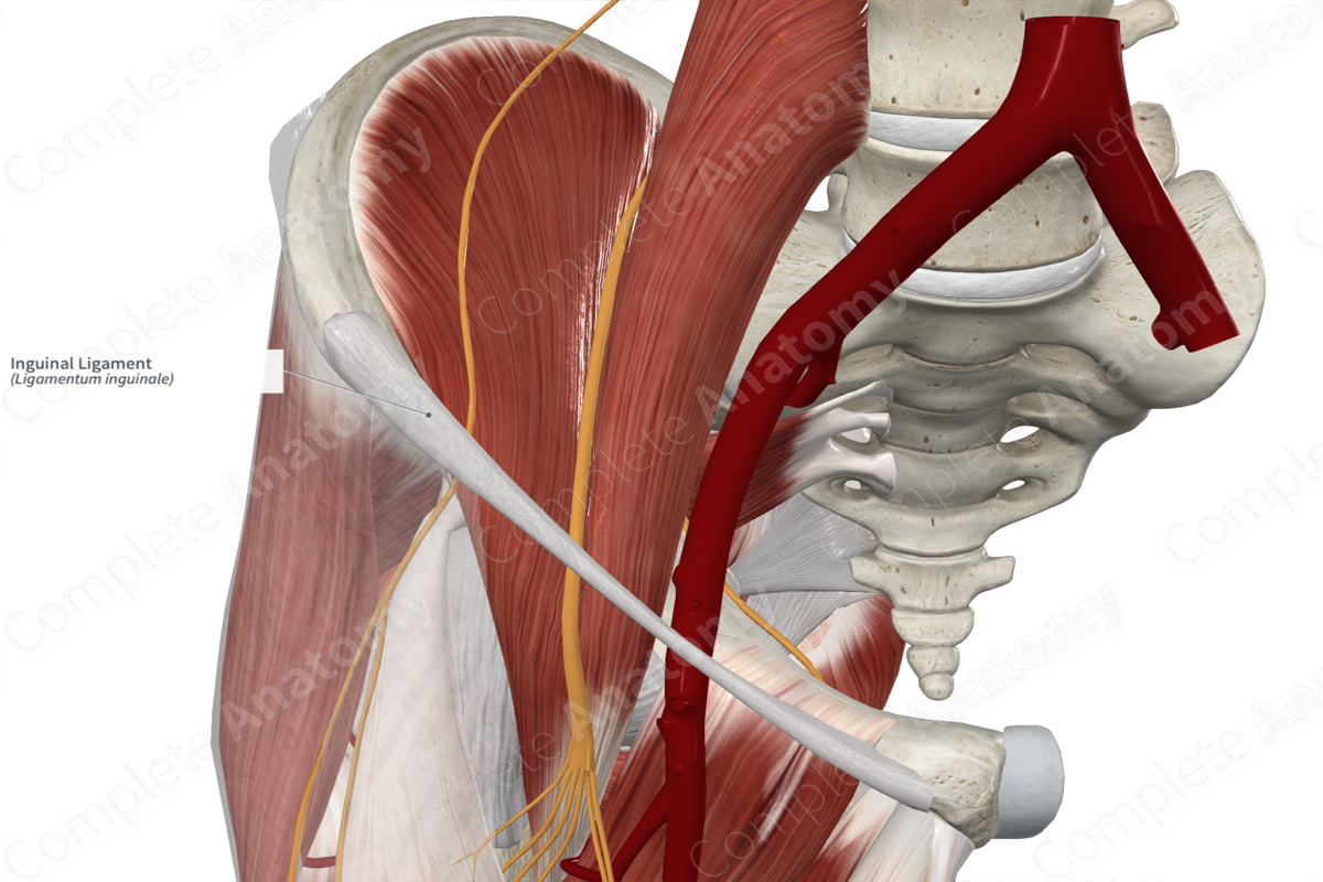
The inguinal ligament runs from the anterior superior iliac spine to the pubic tubercle and along the pectin pubis (Lytle, 1974). The inguinal ligament forms the inferior border of the inguinal (Hesselbach’s) triangle, while the rectus abdominis muscle and inferior epigastric vessels mark the medial and lateral borders (Dorland, 2011).
The external iliac vessels, femoral nerve, and the lateral cutaneous nerve of the thigh run deep to the inguinal ligament on their inferior course into the anterior thigh. The saphenous opening in the fascia lata of the thigh lies inferior to the medial part of the inguinal ligament.
Function
The inguinal ligament supports to femoral sheath and helps in holding open the thin walled femoral vein fixed inside the sheath under the stresses and strains of movement and intra-abdominal pressure changes (Lytle, 1974).
List of Clinical Correlates
- Inguinal hernias
References
Dorland, W. (2011) Dorland's Illustrated Medical Dictionary. 32nd edn. Philadelphia, USA: Elsevier Saunders.
Lytle, W. J. (1974) 'The inguinal and lacunar ligaments', J Anat, 118(Pt 2), pp. 241-51.
Learn more about this topic from other Elsevier products

