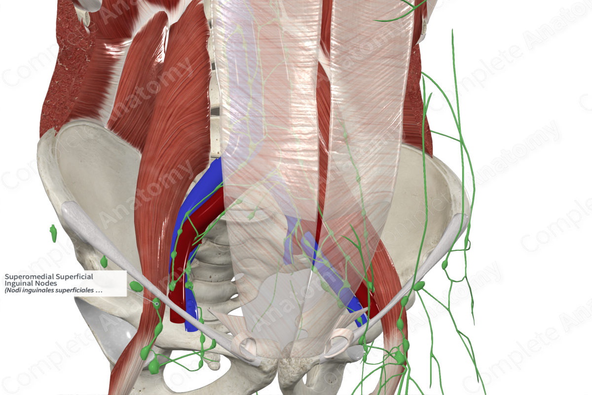
Superomedial Superficial Inguinal Nodes
Nodi inguinales superficiales superomediales
Read moreQuick Facts
Location: Near the termination of the superficial epigastric and external pudendal veins.
Drainage: Superficial layers of anterior abdominal wall (inferior to umbilicus), external genitalia, and perineum.
Direction of Flow: External iliac lymph nodes and deep inguinal lymph nodes > common iliac lymph nodes > lateral aortic lymph nodes (left) and lateral caval lymph nodes (right) > left and right lumbar lymph trunk > cisterna chyli > thoracic duct.


Related parts of the anatomy
Description
The superomedial superficial inguinal lymph nodes usually contain two nodes that are located near the terminal ends of the superficial epigastric and external pudendal veins.
This group of nodes receive lymphatic drainage from the anterior abdominal wall, inferior to the umbilicus, the external genitalia, perineum, anus, lower third of the vagina, and uterine horns in females or the skin of the scrotum in males.
Their efferent vessels pass to the external iliac nodes, with some intercalated vessels passing to the deep inguinal lymph nodes.
Learn more about this topic from other Elsevier products

