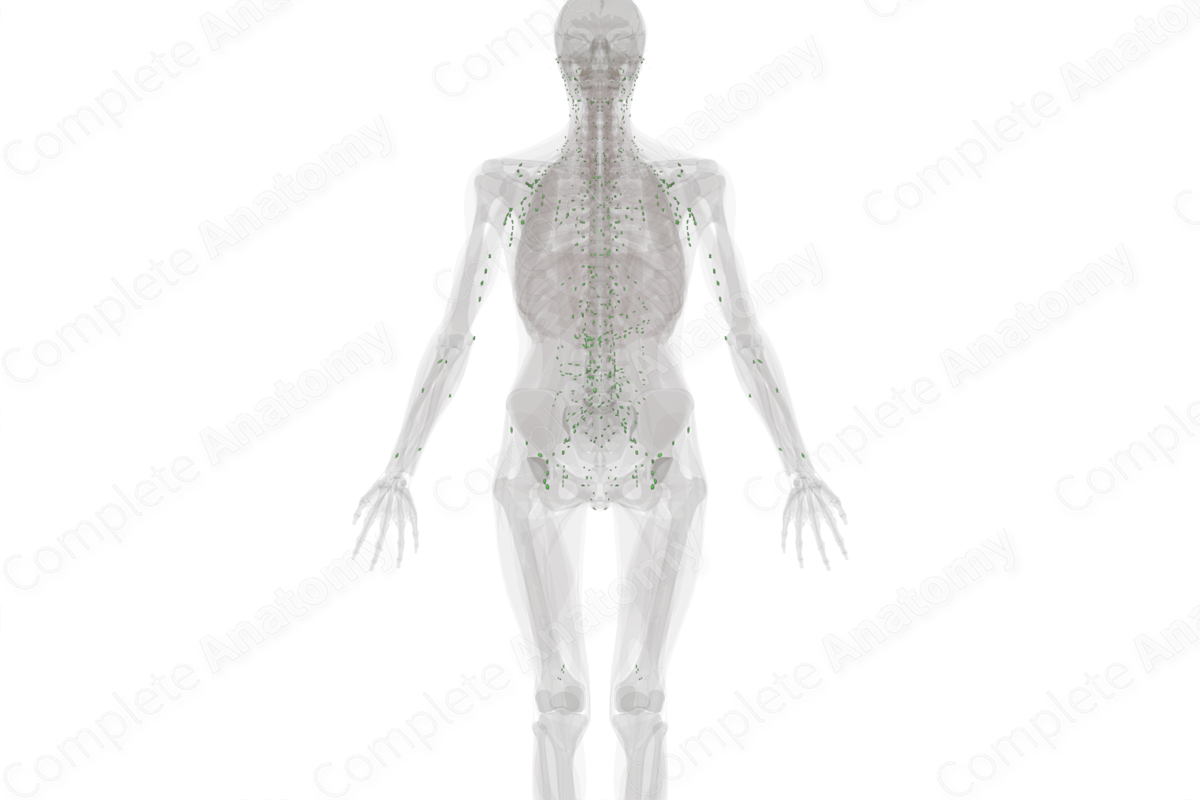
Structure
Lymph nodes are specialized secondary organs of the lymphatic system, resembling kidneys in shape. They range between a few millimeters, and a few centimeters in length.
Structurally, lymph nodes comprise an inner medulla and an outer cortex which are encased in a fibrous capsule. Folds of the capsule invade the medulla to form structures known as trabeculae. There is a space between the capsule and the outer cortex called the subcapsular sinus which allows for the free movement of the lymph (Standring, 2016).
The substance of the nodes is divided into separate compartments known as nodules, or lobules, due to the invaginating trabeculae. A reticulin meshwork provides structural support to the matrix within the node. Additionally, the matrix provides an anchor point for macrophages, lymphocytes, and dendritic cells, and renders the growth and regulatory factors necessary for activation and maturation of immune cells.
Within these lymph nodes is a network of small endothelial-lined channels known as sinuses comprising of the subcapsular sinus and the trabecular and medullary sinuses. The afferent lymphatic vessels drain lymph into the subcapsular sinus. The lymphatic fluid then drains into the trabecular and medullary sinuses, and then to the efferent lymph vessels where it leaves the node at the node’s hilum.
The lymph nodes contain two major immune-response cells; the B and the T-lymphocytes. The B cells congregate as lymphoid follicles towards the outer margin of each nodule (superficial cortex), while the T cells are located mainly in the paracortex, just deep to the superficial cortex (Standring, 2016).


Function
The lymph node is responsible for receiving lymph drained by afferent lymph vessels from surrounding tissue. The lymph node filters the lymph and antigenic material becomes trapped and phagocytosed by the macrophages. It is also the site where B and T cell lymphocytes proliferate and mature.
References
Standring, S. (2016) Gray's Anatomy: The Anatomical Basis of Clinical Practice. Gray's Anatomy Series: Elsevier Limited.
Learn more about this topic from other Elsevier products
Lymph node histology: Video, Causes, & Meaning

Lymph node histology: Symptoms, Causes, Videos & Quizzes | Learn Fast for Better Retention!

