Quick Facts
Origin: Maxillary nerve.
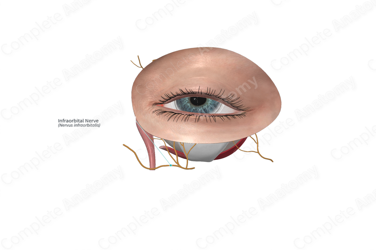
Course: Passes anteriorly from the pterygopalatine fossa into the orbit through the inferior orbital fissure. Travels within the inferior orbital groove along the floor of the orbit. It exits the orbit passing through the infraorbital foramen just below the lower eyelid and onto the cheek.
Branches: Anterior and middle superior alveolar branches, inferior palpebral nerves, external and internal nasal and superior labial branches.
Supply: Conveys general sensory information from the skin of the anterior face overlying the maxilla and the anterior cheek, the upper lip, lateral portions of the nose, and mucosa of the upper lip, anterior-most nasal cavity, and maxillary sinus.


Origin
The infraorbital nerve originates as one of the major branches of the maxillary nerve within the pterygopalatine fossa. It is often considered the anterior continuation of the maxillary nerve.
Course
The infraorbital nerve exits the pterygopalatine fossa by passing anteriorly through the inferior orbit fissure. It continues anteriorly along the floor of the orbit in the infraorbital groove. At variable position along its course, the infraorbital groove becomes enclosed superiorly by bone, thus becoming the infraorbital canal. This canal opens distally at the infraorbital foramen of the maxilla where the nerve emerges onto the anterior face, roughly 5-10 mm below the inferior margin of the orbit, lateral to the pyriform aperture of the nasal cavity.
Branches
The infraorbital nerve gives rise to five branches.
—The middle and anterior superior alveolar nerves are both given off within the infraorbital groove/canal.
—The inferior palpebral branches form upon emerging from the infraorbital foramen and immediately ascend, passing deep to orbicularis oculi muscle into the lower eyelid
—The nasal branches of the infraorbital nerve also form upon emerging from the infraorbital foramen and immediately pass medially to the lateral surface of the nose.
—The superior labial branches also form upon emerging from the infraorbital foramen and immediately pass inferiorly, deep to levator labii superioris muscle.
Supplied Structures
The middle and anterior superior alveolar nerves supply the mucosa of the maxillary sinus, as well as the first molar and premolar, and the canine and incisor, upper teeth, respectively. The inferior palpebral nerve supplies the skin of the upper cheek and lower eyelid including the conjunctiva on its inner surface. The nasal branches supply the skin of the medial cheek and lateral nose including the mucosa on its inner surface. The superior labial branches supply the skin of the lower cheek, upper lip (including the philtrum), and vermillion zone, as well as the mucosa of its buccal surface.
The maxillary nerve and its infraorbital branch is properly considered a purely general sensory nerve. However, within the pterygopalatine fossa is housed a major parasympathetic ganglion, the pterygopalatine ganglion, whose postganglionic fibers opportunistically join the branches of the maxillary nerve and follow them to their smooth muscle and glandular targets in the periphery. Those that hitch a ride with the infraorbital nerve innervate the labial oral mucus glands of the upper lip and mucosa.
List of Clinical Correlates
—Trigeminal neuralgia
—Maxillary sinusitis
—Abscessed upper tooth
