Quick Facts
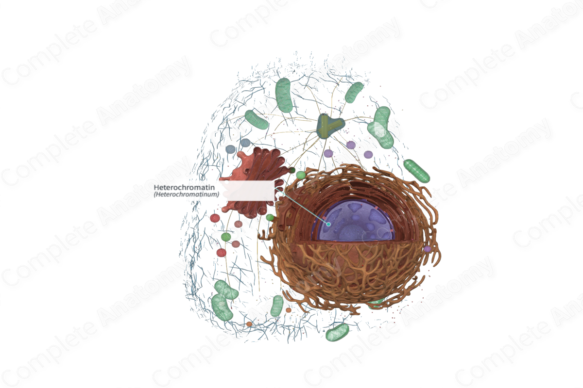
Heterochromatin is the form of chromatin that is darkly staining and tightly coiled in interphase; it is composed of repetitive DNA, is late to replicate, and is transcriptionally inactive (Dorland, 2011).
Related parts of the anatomy
Structure and/or Key Feature(s)
Compact or very condensed chromatin visible with the light microscope is called heterochromatin. It's more tightly packed DNA.
A clump of chromatin (heterochromatin) frequently seen in female cells is called sex chromatin and is one of the two X chromosomes and is generally inactive.
Heterochromatin stains with basic dyes and hematoxylin (Ross and Pawlina, 2006; Ovalle, Nahirney and Netter, 2013; McKinley, O'Loughlin and Pennefather-O'Brien, 2016).
Function
Heterochromatin is prominent in cells that are metabolically quiescent.
References
Dorland, W. (2011) Dorland's Illustrated Medical Dictionary. 32nd edn. Philadelphia, USA: Elsevier Saunders.
McKinley, M. P., O'Loughlin, V. D. and Pennefather-O'Brien, E. E. (2016) Human Anatomy. 5th edn.: McGraw-Hill Education.
Ovalle, W. K., Nahirney, P. C. and Netter, F. H. (2013) Netter's Essential Histology. ClinicalKey 2012: Elsevier Saunders.
Ross, M. H. and Pawlina, W. (2006) Histology: A text and atlas. Lippincott Williams & Wilkins.
