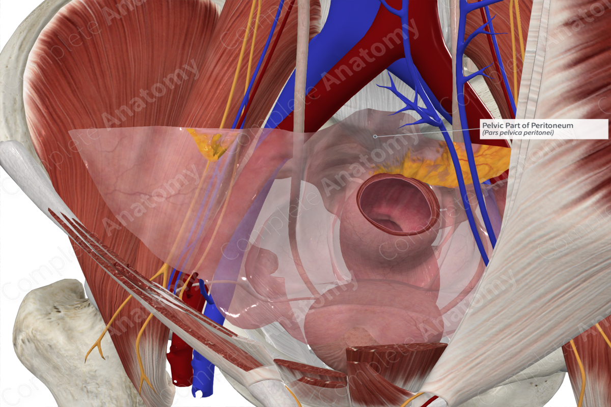
Structure
The peritoneum is a complex, continuous serous membrane consisting of a layer of mesothelium and varying degrees of connective and adipose tissue. Visually, it’s largely unremarkable, smooth, and has a lubricated surface due to the presence of peritoneal fluid.
Anatomical Relations
The urogenital peritoneum is the extension of the parietal abdominal peritoneum inferiorly where it’s reflected over the urogenital organs. Inferiorly, the pelvic organs are situated amongst endopelvic fascia, often contributing to the peritoneal folds and depressions.
The urogenital peritoneum does not fully cover the urogenital organs, but drapes over them; therefore, the organs are not considered intraperitoneal (rather subperitoneal), except for the uterine tubes and ovaries in females (Standring, 2016). The urogenital peritoneum in males remains a closed membranous sac, however, in females, there is a small ostium at the distal end of each uterine tube allowing entry of an ovum into the tube.
Function
The peritoneum secretes peritoneal fluid which helps lubricate viscera within the abdominal and pelvic cavities, reducing friction between organs. This is especially true of dynamic organs involved in peristalsis or those that distend due to changes of volume, such as the bladder. Secondly, the peritoneum aids in the immune response as the peritoneal fluid contains various immune cells. The peritoneal fluid actively flows around the peritoneal cavity and is eventually absorbed by lymphatics. The peritoneum, like other tissues, responds to trauma and inflammation, thus helping to protect the viscera (Moore, Dalley and Agur, 2013).
List of Clinical Correlates
- Peritonitis
- Ascites
- Adhesions
References
Moore, K. L., Dalley, A. F. and Agur, A. M. R. (2013) Clinically Oriented Anatomy. Clinically Oriented Anatomy 7th edn.: Wolters Kluwer Health/Lippincott Williams & Wilkins.
Standring, S. (2016) Gray's Anatomy: The Anatomical Basis of Clinical Practice. Gray's Anatomy Series 41 edn.: Elsevier Limited.
Learn more about this topic from other Elsevier products


