Quick Facts
Location: Within the true pelvis, against the anterior surface of the sacrum, from the level of S3 vertebra to the pelvic floor.
Arterial Supply: Superior, middle, and inferior anorectal arteries.
Venous Drainage: Superior, middle, and inferior anorectal veins.
Innervation: Parasympathetic: pelvic splanchnic nerves (S2-S4), superior hypogastric, inferior mesenteric, rectal, iliac, coccygeal, and sacral plexuses; Sympathetic: lumbar splanchnic nerves, superior and inferior mesenteric plexus, and inferior hypogastric plexus; visceral afferents (spinal ganglia S2-S4).
Lymphatic Drainage: Inferior mesenteric and external and internal iliac lymph nodes.
Structure/Morphology
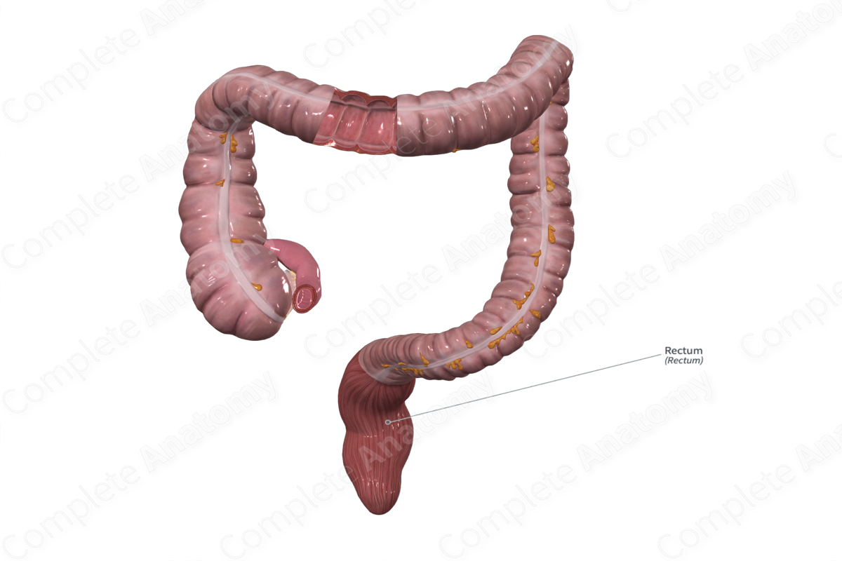
The rectum is a distal continuation of the sigmoid colon that transitions at the level of S3 and extends as far as puborectalis muscle, a component of levator ani that constitutes much of the muscular pelvic floor. Its length is variable, but it's approximately 15 cm (Standring, 2016). Proximally, it lies on the median plane where, its diameter is similar to the sigmoid colon. It dilates and becomes the rectal ampulla.
The rectum follows the concavity of the sacrum and coccyx, curving first posteriorly, then anteriorly as it passes inferiorly. The rectum makes three lateral curves as it descends, from superior to inferior: to the right, to the left (most prominent) and to the right again. Its distal end returns to the midline.
The anteriorly directed distal rectum lies in contrast to the posteriorly directed proximal anal canal. Thus, an angle, the anorectal angle, forms at the anorectal junction. It is maintained by the “muscular sling” of the puboanalis (puborectalis) muscle. The anorectal junction is just inferior to the distal tip of the coccyx, approximately 2–3 cm anterior to it (Standring, 2016). In males, this corresponds to the apex of the prostate.
Externally, the rectum does not contain sacculations or omental appendices which distinguish it from the colon. Its longitudinal muscle layer is continuous as the distinct teniae coli fan out and merge just proximal to the rectosigmoidal junction.
Internally, the inner mucosal surface has several longitudinal folds in the distal rectum that disappear when the rectum is distended. There are also usually three transverse rectal folds, and these become prominent during distension.
Anatomical Relations
Peritoneum covers the anterior and lateral aspects of the upper one third of the rectum and the anterior aspect of the middle third. The peritoneal attachment is relatively loose allowing for expansion of the rectum. The lower one third of the rectum is infraperitoneal.
At the rectal ampulla, a few longitudinal muscle fibers pass forward from the anterior rectal wall to the perineal body and urethra (rectourethral or puborectalis muscles).
Posteriorly, the rectum is related to the presacral fascia, the lower three sacral vertebrae, coccyx, median and lateral sacral arteries and veins, and the sacral sympathetic chain.
Anterolateral to the proximal rectum on the left-hand side is the sigmoid colon, superior to the rectum is the ileum, and inferior is the puboanalis muscle.
In males, the rectum is separated from the posterior aspect of the bladder by the rectovesical pouch. In females, the imposition of the uterus makes the corresponding space the rectouterine pouch (of Douglas).
Function
The rectal ampulla acts as a temporary storage site for feces before defecation via the anal canal.
Arterial Supply
The superior, middle, and inferior anorectal arteries supply the rectum (Standring, 2016).
The superior anorectal artery is the continuation of the inferior mesenteric artery. It passes directly inferior, crossing the pelvic brim to enter the mesorectal fascia (or mesorectum) at the level of S3 vertebra. Here it divides into two posterolateral branches that shift laterally as they travel inferiorly, eventually entering the rectal wall and anastomosing with branches of the middle and inferior anorectal arteries.
The middle anorectal arteries originate from the anterior division of the internal iliac artery. They travel anterolaterally in the mesorectum and give off several branches that pierce the walls of the rectum. They aid in the vascular supply for the middle one third of the rectum.
The inferior anorectal arteries arise from the internal pudendal arteries in the posterior portion of the pudendal canal. The inferior rectal arteries travel anteromedially in the horizontal plane, passing across the ischioanal fossa to the lateral aspects of the rectum and anal canal and courses inferiorly.
Venous Drainage
Venous drainage of the rectum is completed by the superior, middle, and inferior anorectal veins that follow a similar course to the arteries.
Innervation
Parasympathetic innervation of the rectum is derived from many sources, including the pelvic splanchnic nerves (S2-S4), the superior hypogastric, inferior mesenteric, rectal, iliac, coccygeal and sacral plexuses.
Sympathetic innervation arises from lumbar splanchnic nerves (L1-L2) and synapse in inferior mesenteric plexus while lumbar and sacral splanchnic nerves synapse in the superior and inferior hypogastric, rectal and iliac plexuses.
All visceral afferents travel with parasympathetic nerves to S2-S4 sensory ganglia (Standring, 2016).
Lymphatic Drainage
The superior rectal lymph nodes follow the superior rectal artery and drain into the inferior mesenteric nodes. A portion of the distal rectum is drained by the Pararectal lymph nodes, which return lymph to the external and internal iliac lymph nodes (Földi et al., 2012).
List of Clinical Correlates
- Anorectal incontinence
References
Földi, M., Földi, E., Strößenreuther, R. and Kubik, S. (2012) Földi's Textbook of Lymphology: for Physicians and Lymphedema Therapists. Elsevier Health Sciences.
Standring, S. (2016) Gray's Anatomy: The Anatomical Basis of Clinical Practice. Gray's Anatomy Series 41st edn.: Elsevier Limited.
Learn more about this topic from other Elsevier products


