Quick Facts
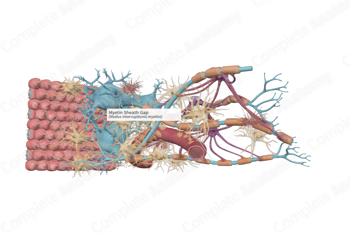
The myelin sheath gap, or node of Ranvier, are the constrictions occurring on myelinated nerve fibers at regular intervals of about 1 mm; at these sites the myelin sheath is absent and the axon is enclosed only by Schwann cell processes (Dorland, 2011).
Related parts of the anatomy
Structure and/or Key Features
The myelin sheath is produced by oligodendrocytes in the central nervous system and by Schwann cells in the peripheral nervous system.
Oligodendrocytes are small cells and extend long processes towards neighboring axons. Each process wraps around a portion of the same axon or several axons. The myelin sheath that wraps around the axon becomes segmented at regular intervals by the nodes of Ranvier, or myelin sheath gaps.
The area between adjacent nodes of Ranvier is known as the internode. The length of the internode is correlated with the diameter of the axon (Ross and Pawlina, 2006).
Anatomical Relations
Nodes of Ranvier are located in the intervals between adjacent myelin sheaths. “Foot processes” extending from the astrocyte cell body can be found in large numbers at the nodes of Ranvier (Splittgerber, 2018).
Function
The nodes of Ranvier facilitate the rapid conduction of nerve impulses. Sodium channels embedded in the axolemma are greatest in density at the nodes of Ranvier and lowest in density at the internodes (Standring, 2016). The high concentration of sodium channels at the nodes facilitates “saltatory conduction” which is the process by which the action potential is propagated from node to node very rapidly along myelinated nerve fibers.
Clinical Correlates
Deficiencies in any of the oligodendrocytes myelin-specific proteins seem related to demyelinating diseases of the central nervous system. Multiple sclerosis is characterized by demyelination in white matter of the CNS. The degenerated myelin sheaths are removed by microglial cells and then astrocytes proliferate to form a gliotic scar (Splittgerber, 2018).
References
Dorland, W. (2011) Dorland's Illustrated Medical Dictionary. 32nd edn. Philadelphia, USA: Elsevier Saunders.
Ross, M. H. and Pawlina, W. (2006) Histology: A text and atlas. Lippincott Williams & Wilkins.
Splittgerber, R. (2018) Snell's Clinical Neuroanatomy. Wolters Kluwer Health.
Standring, S. (2016) Gray's Anatomy: The Anatomical Basis of Clinical Practice. Gray's Anatomy Series 41 edn.: Elsevier Limited.
