Quick Facts
Ependymal cells make up the ependyma, the lining membrane of the ventricles of the brain and of the central canal of the spinal cord (Dorland, 2011).
Related parts of the anatomy
Structure and/or Key Features
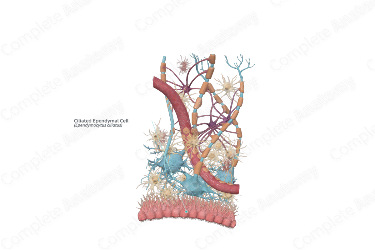
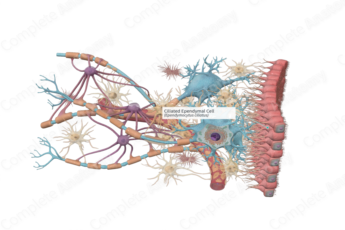
Ependymal cells (ependymocytes) are epithelial-like glial cells that line the ventricles of the brain and the central canal of the spinal cord. These cells are either columnar or cuboidal cells and, in some areas of the central nervous system (CNS), exhibit cilia on their apical surface. Each ependymal cell has around 20 central apical cilia each of which is surrounded by short microvilli (Standring, 2016).
Ependymal cells lack an external lamina and are joined by tight junctional complexes at their apical surfaces. Their basal surfaces have numerous infoldings interdigitating with processes from astrocytes (Ross and Pawlina, 2006).
Ependymal cells are divided into three groups.
—Ependymocytes line the ventricles of the brain and the central canal of the spinal cord and are in contact with cerebrospinal fluid.
—Tanycytes are very specialized and are most numerous in the floor of the fourth ventricle overlying the median eminence of the hypothalamus. They are in contact with cerebrospinal fluid but do not have cilia on their apical surface. However, they do have long basal processes that pass into the neural tissue of the median eminence and place foot-like process on blood vessels.
—Modified ependymal cells are joined by tight junctions and are associated with blood capillaries to form the choroid plexus (Ross and Pawlina, 2006).
Anatomical Relations
The ependymal cells line the ventricles of the brain and the central canal of the spinal cord. They are in contact with the processes of astrocytes, which extend towards the fluid-filled spaces of the central nervous system (Ross and Pawlina, 2006).
Function
The cilia on the apical surfaces of the ependymal cells aid in the circulation of the cerebrospinal fluid, while microvilli assist in absorption of the cerebrospinal fluid.
Tanycytes are involved with transporting substances from the cerebrospinal fluid to the portal circulation of the hypothalamus. They may also monitor circulating metabolites in the cerebrospinal fluid since they appear to be sensitive to glucose concentrations.
The choroid plexus produces cerebrospinal fluid (Ross and Pawlina, 2006).
Clinical Correlates
Ependymomas arise from ependymal cells lining the ventricles particularly in the region of the cerebellum.
References
Dorland, W. (2011) Dorland's Illustrated Medical Dictionary. 32nd edn. Philadelphia, USA: Elsevier Saunders.
Ross, M. H. and Pawlina, W. (2006) Histology: A text and atlas. Lippincott Williams & Wilkins.
Standring, S. (2016) Gray's Anatomy: The Anatomical Basis of Clinical Practice. Gray's Anatomy Series 41 edn.: Elsevier Limited.
