Quick Facts
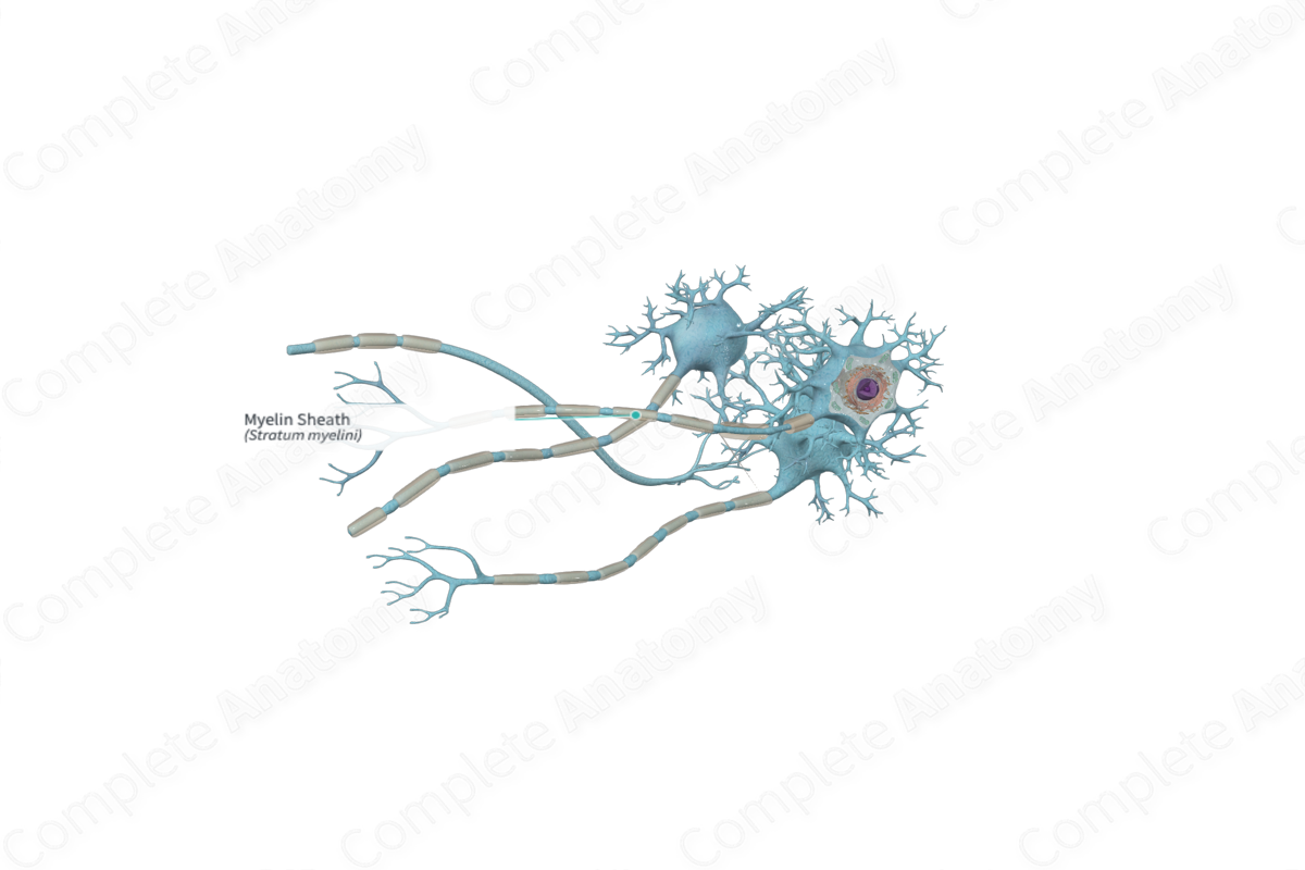
The myelin sheath is the cylindrical covering on the axons of some neurons; it consists of concentric layers of myelin, formed in the peripheral nervous system by the plasma membrane of Schwann cells, and in the central nervous system by oligodendrocytes. It is interrupted at intervals along its length by gaps known as nodes of Ranvier. Myelin is an electrical insulator that serves to speed the conduction of nerve impulses (Dorland, 2011).
Related parts of the anatomy
Structure and/or Key Features
The myelin sheath is produced by oligodendrocytes in the central nervous system (CNS) and by Schwann cells in the peripheral nervous system (PNS). The formation of the myelin sheath is more complex in the CNS than it is in the PNS. Oligodendrocytes produce long processes that extend towards an axon and wraps its plasmalemma around the axon in concentric layers. In some regions of the CNS, oligodendrocytes can enclose up to fifty axons in separate myelin sheaths (Mancall and Brock, 2011). The area of the axon covered by the myelin sheath is called the internodal segments. The thickness of the myelin sheath during the process of myelination is determined by the axon diameter (Ross and Pawlina, 2006).
Myelin is a whitish lipoprotein complex that consists predominantly of lipid bilayers and membrane proteins. The high lipid content is partly removed by traditional histological processes leaving a characteristic “halo” around the axon (Mescher, 2013). The myelin sheath in the CNS differs from the myelin sheath in the PNS. This is because oligodendrocytes and Schwann cells express different myelin-specific proteins during myelination.
Anatomical Relations
The myelin sheath extends from the initial segment of the axon until right before its terminal branches.
Function
The myelin sheath forms an insulating layer around the axon and prevents leakage of the electrical current from the axon. In addition, the myelin sheath increases the speed by which nerve impulses are conducted along the axon and makes the propagation of nerve impulses more energy efficient (Marieb, Wilhelm and Mallatt, 2012).
The myelin sheath also functions to maintain an ideal ionic environment that is suitable for the conduction and propagation of the action potentials (Mescher, 2013).
Clinical Correlates
Demyelinating diseases preferentially damage the myelin sheath and so result in decreased or loss of transmission of nerve impulses along the nerve fiber. In multiple sclerosis, the myelin sheath is destroyed.
References
Dorland, W. (2011) Dorland's Illustrated Medical Dictionary. 32nd edn. Philadelphia, USA: Elsevier Saunders.
Mancall, E. L. and Brock, D. G. (2011) Gray's Clinical Neuroanatomy: The Anatomic Basis for Clinical Neuroscience. Elsevier Health Sciences.
Marieb, E. N., Wilhelm, P. B. and Mallatt, J. (2012) Human Anatomy. fifth edn.: Benjamin Cummings.
Mescher, A. (2013) Junqueira's Basic Histology: Text and Atlas. 13th edn.: McGraw-Hill Education.
Ross, M. H. and Pawlina, W. (2006) Histology: A text and atlas. Lippincott Williams & Wilkins.
