Morphology/Structure
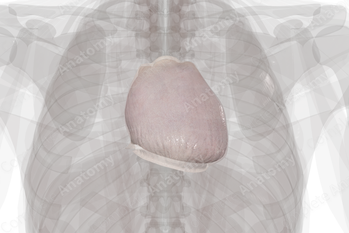
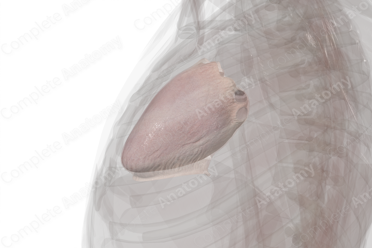
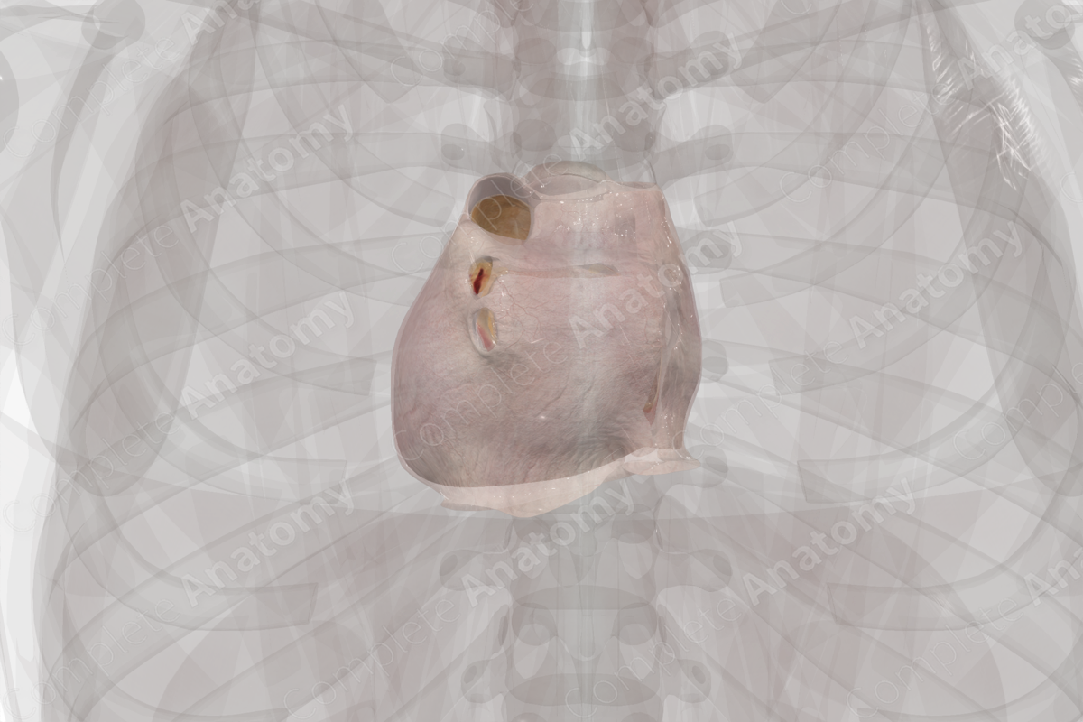
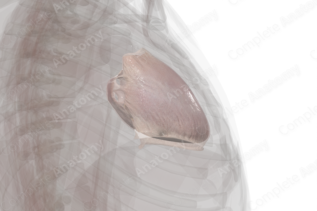
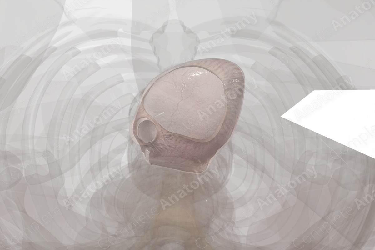
The pericardium envelops the heart and the base of the great vessels. It consists of an outer fibrous layer and two inner serous pericardial layers. The fibrous layer is attached to the sternum anteriorly, the central tendon of the diaphragm inferiorly, the vertebral column posteriorly, the mediastinal pleural laterally, and to the outer layer of the base of the great vessels. However, it’s not attached to the heart itself.
There are two layers of serous pericardium, an outer parietal layer that attaches to the internal surface of the fibrous layer of pericardium; and an inner visceral layer, which attaches to the heart as the epicardium. Between the two serous layers is a potential space called the pericardial sac, which contains serous fluid.
Key Features/Anatomical Relations
The pericardium occupies the middle compartment of the inferior mediastinum and sits between the lungs. The sternum is placed anteriorly, structures of the posterior mediastinum (the thoracic duct, vagus nerves, esophagus, and descending aorta) sit behind the pericardium; and the lungs and mediastinal pleura sit laterally, while the diaphragm sits inferiorly.
The pericardiacophrenic vessels that accompany the phrenic nerve within the fibrous layer of pericardium provide the bulk arterial supply and venous drainage of the pericardium. Additionally, the phrenic nerve provides sensory innervation to the fibrous and parietal serous layers of pericardium. Pain is typically felt substernal but may radiate from the upper border of the trapezius muscle. The visceral layer of serous pericardium is innervated by the sympathetic (aortic and cardiac plexuses) and parasympathetic fibers from the vagus nerve (CN X); thus, this layer is insensitive to pain.
Function
The fibrous layer of pericardium ensures stability by attaching to surrounding structures namely the sternum, vertebral column, and diaphragm. It also provides a physical limit to cardiac distension and a barrier to the spread of infection. The serous layer produces fluid, which enables friction-free movement of the heart within the pericardium.
List of Clinical Correlates
- Cardiac tamponade
- Pericardial effusion
- Pericarditis
- Pericardial rub
Learn more about this topic from other Elsevier products

