Quick Facts
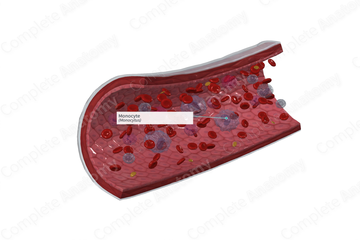
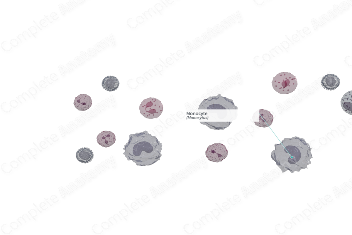
A monocyte is a mononuclear phagocytic leukocyte, 13 to 25 μm in diameter, with an ovoid or kidney-shaped nucleus, containing lacy, linear chromatin and abundant gray-blue cytoplasm filled with fine reddish and azurophilic granules. Formed in the bone marrow from promonocytes, monocytes are transported to tissues such as the lung and liver, where they develop into macrophages (Dorland, 2011).
Related parts of the anatomy
Cell Morphology
Monocytes are the largest of the leukocytes, with a diameter of 14–24 μm, and account for 4–8% of leukocytes (100–700 monocytes per microliter of blood) (Marieb, Wilhelm and Mallatt, 2012). Monocytes are larger than lymphocytes and are recognizable from their indented horseshoe or kidney-shaped nucleus. The nuclear chromatin of monocytes isn’t as condensed as it is in lymphocytes and monocytes possess more cytoplasm than lymphocytes. The cytoplasm of monocytes may contain some small dispersed granules, typically lysosomes. However, since monocytes lack obvious granules they are classified as agranulocytes. Monocytes have a well-developed Golgi apparatus, smooth endoplasmic reticulum, rough endoplasmic reticulum, and small mitochondria.
Production
In the red bone marrow, hematopoietic stem cells differentiate into myeloid stem cells which give rise to monocytes. Monocytes travel from the bone marrow and circulate in the blood for around 3 days. They then enter body tissues and differentiate into various types of phagocytic cells. Development of monocytes takes 2–3 days (Marieb, Wilhelm and Mallatt, 2012).
Monocytes can differentiate into macrophages found in the lymph nodes, liver, spleen, alveoli, and connective tissue (Pawlina, 2016).
Function
When monocytes transform into macrophages, they function as antigen presenting cells. That means that they bind antigens and present it to T lymphocytes for recognition and elimination. Macrophages move via an ameboid movement and phagocytose bacteria, other cells, molecules, and tissue debris (Pawlina, 2016; Marieb, Wilhelm and Mallatt, 2012).
List of Clinical Correlates
—Monocytosis
—Monocytopenia
References
Dorland, W. (2011) Dorland's Illustrated Medical Dictionary. 32nd edn. Philadelphia, USA: Elsevier Saunders.
Eroschenko, V. P. (2008) DiFiore's Atlas of Histology with Functional Correlations. Wolters Kluwer Health/Lippincott Williams & Wilkins.
Marieb, E. N., Wilhelm, P. B. and Mallatt, J. (2012) Human Anatomy. 14th edn.: Benjamin Cummings.
Pawlina, W. 2016. Histology: A text and atlas with correlated cell and molecular biology. 7th ed. Philadelphia: Wolters Kluwer.
