Quick Facts
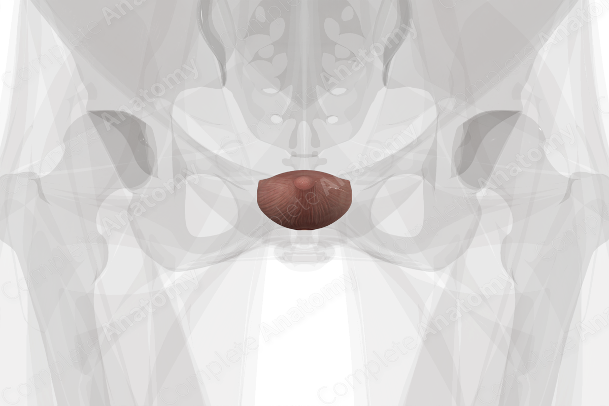
Location: Base of the pelvis.
Arterial Supply: Superior and inferior vesical arteries.
Venous Drainage: Superior (and inferior) vesical veins.
Innervation: Via the vesical and hypogastric plexus: Sympathetic: sacral splanchnic nerves; Parasympathetic: pelvic splanchnic nerves; visceral afferent fibers.
Lymphatic Drainage: Paravesical lymph nodes.
Related parts of the anatomy
Structure/Morphology
The urinary bladder is essentially a hollow, muscular reservoir. The morphology changes as it becomes filled with urine, moving a portion of the bladder into the greater pelvis.
Anatomical Relations
The urinary bladder is directly posterior to the pubic symphysis and superior to the pelvic diaphragm. In females, the uterus, cervix, and vagina are posterior to the bladder. In males, the rectum is posterior to the bladder.
Function
The urinary bladder stores urine that has passed from the kidneys through the ureters. Urination occurs when the internal urethral sphincter (distal portion of the bladder) relaxes and the detrusor (smooth muscle) of the bladder contracts, expelling urine into the urethra.
Arterial Supply
The urinary bladder receives a dual blood supply that supplies the superior and inferior parts of the organ. Superior vesical arteries are branches off the umbilical artery. In males, the inferior vesical arteries branch directly from the internal iliac arteries. In females, the vaginal branch of the uterine artery serves the function of the inferior vesical and supplies the inferior bladder (Tubbs et al., 2016).
Venous Drainage
The bladder is drained by the vesical venous plexus, where blood is returned to the internal iliac vein via the superior (and in the male, the inferior) vesical veins.
Innervation
The nerves supplying the bladder arise from the pelvic plexuses. Sympathetic fibers are derived from the sacral splanchnic nerve and synapse with the postganglionic neuronal cell bodies inside the celiac and mesenteric plexuses. From here the hypogastric plexuses descend into the pelvis and connect with the vesical plexus surrounding the bladder.
Parasympathetic fibers arise from the neuronal cell bodies in the pelvic splanchnic nerve.
Vesical afferent nerves are concerned with pain and awareness of distension of the bladder (Burnstock, 1990).
Lymphatic Drainage
The paravesical lymph nodes are found in the subperitoneal tissue surrounding the urinary bladder. These nodes drain to the external (mostly) and internal (minimally) iliac lymph nodes, returning lymph to the lateral aortic and caval lymph nodes and the left and right lumbar lymph trunks (Földi et al., 2012).
List of Clinical Correlates
—Bladder cancer
References
Burnstock, G. (1990) 'Innervation of bladder and bowel', Ciba Found Symp, 151, pp. 2-18.
Földi, M., Földi, E., Strößenreuther, R. and Kubik, S. (2012) Földi's Textbook of Lymphology: for Physicians and Lymphedema Therapists. Elsevier Health Sciences.
Tubbs, R. S., Shoja, M. M. and Loukas, M. (2016) Bergman's Comprehensive Encyclopedia of Human Anatomic Variation. Wiley.
Learn more about this topic from other Elsevier products
Ureter, bladder and urethra histology: Video, Causes, & Meaning

Ureter, bladder and urethra histology: Symptoms, Causes, Videos & Quizzes | Learn Fast for Better Retention!


