Quick Facts
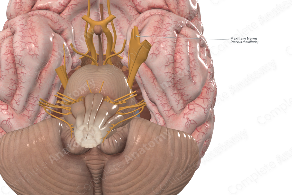
Origin: Trigeminal ganglion.
Course: Runs anteriorly through the cavernous sinus and passes into the pterygopalatine fossa via the foramen rotundum.
Branches: Meningeal branch, infraorbital, zygomatic, and posterior superior alveolar nerves, and branches to the pterygopalatine ganglion. Additionally, there are six branches of the maxillary nerve that originate from the pterygopalatine ganglion, including the orbital branches, pharyngeal nerve, posterior superior nasal branches, greater and lesser palatine nerves, and nasopalatine nerve.
Supply: Conveys general sensation from the skin of the middle face, portions of the nose and nasal cavity, the orbit and sphenoid sinus, the maxillary sinus, and the maxillary teeth.


Related parts of the anatomy
Origin
The maxillary nerve originates at the trigeminal ganglion as the middle of three branches of the trigeminal nerve. It projects anteriorly just inferior to the ophthalmic nerve.
The neurons that make up the maxillary nerve are found in the trigeminal ganglion. They project to the main sensory nucleus of the trigeminal nerve and the spinal nucleus of the trigeminal nerve, both in the pons.
Course
The maxillary nerve emerges from the anterior portion of the trigeminal ganglion, just inferior to the ophthalmic nerve. It runs anteriorly, passing through the lateral portion of the cavernous sinus until it reaches the foramen rotundum. It passes through the foramen rotundum to enter the upper pterygopalatine fossa.
At this point it gives off most of its direct branches, with the infraorbital nerve representing the anterior continuation of the maxillary nerve. Fibers descend to and through the pterygopalatine ganglion to give off additional branches (Norton, 2016).
Branches
The maxillary nerve gives rise to five branches directly and six branches indirectly via the pterygopalatine ganglion.
The direct branches include the:
meningeal branch;
zygomatic nerve;
infraorbital nerve;
posterior superior alveolar nerve;
branches to pterygopalatine ganglion.
The branches to the pterygopalatine ganglion convey provide indirect branches of the maxillary nerve. They transmit general sensation as well as parasympathetic fibers from the pterygopalatine ganglion to the zygomatic nerve. These indirect branches of the maxillary nerve include:
orbital branches;
pharyngeal nerve;
posterior superior nasal nerves;
greater palatine nerve;
lesser palatine nerve;
nasopalatine nerve.
Supplied Structures
The maxillary nerve conveys general sensation from portions of the skin and mucosal linings of the middle face, orbit, paranasal sinuses, nasal cavity, palate, and pharynx. Some of the nerves that branch off the maxillary nerve pick up parasympathetic fibers, but the maxillary nerve itself does not have any.
The meningeal branch of maxillary nerve conveys general sensation from much of the cranial dura mater, particularly that of the middle cranial fossa.
The zygomatic nerve conveys sensation from the skin overlying the zygomatic bone and arch. The zygomatic nerve also carries postganglionic parasympathetic fibers that synapse in the pterygopalatine ganglion with the preganglionic greater petrosal nerve. These fibers run with the zygomatic nerve in the orbit until they communicate with the lacrimal nerve to innervate the lacrimal gland.
The infraorbital nerve runs anteriorly into the lower portion of the orbit. It continues forward through the infraorbital foramen to innervate the skin of the face overlying the maxilla and the upper lips. Branches of the infraorbital nerve also convey sensation from the maxillary sinus and anterior teeth of the maxilla (incisors, canines, and premolars). The posterior superior alveolar nerve innervates the posterior maxillary teeth, specifically the molars.
There are typically two ganglionic branches which run inferiorly to the pterygopalatine ganglion. All sensation from the indirect branches passes through the ganglion and the ganglionic branches to reach the maxillary nerve. These fibers do not synapse in the pterygopalatine ganglion. They include sensory fibers from the nasal cavity, the palate, ethmoid air cells, and the nasopharynx. The ganglionic branches also transmit parasympathetic fibers, destined for the lacrimal gland, to the maxillary nerve and its direct branches. These parasympathetic fibers originate in the pterygopalatine ganglion where they synapse with the preganglionic parasympathetic fibers of the nerve of the pterygoid canal (Norton, 2016).
The orbital branches are small and innervate portions of the sphenoid sinus and the orbital periosteum. The pharyngeal nerve runs posteriorly through the palatovaginal canal and innervates the mucosa of the nasopharynx.
The posterior superior nasal nerves run anteriorly and innervate mucosa overlying the posterior and superior portions of both the nasal concha and septum, as well as some posterior ethmoid air cells.
The greater palatine nerve descends, giving off the posterior inferior nasal branches to the mucosa of the inferior concha. After passing through the greater palatine foramen, the greater palatine nerve spreads to convey sensory information from most of the mucosa of the hard palate and the lingual gingiva.
The lesser palatine nerve descends through the lesser palatine foramen to innervate the mucosa of the soft palate, uvula, and palatine tonsil.
The nasopalatine nerve runs medially through the sphenopalatine foramen to innervate the inferior nasal septum and, after passing through the incisive canal, the anterior hard palate and lingual gingiva.
References
Norton, N. S. (2016) Netter's Head and Neck Anatomy for Dentistry E-Book. Elsevier Health Sciences.
Learn more about this topic from other Elsevier products

