Description
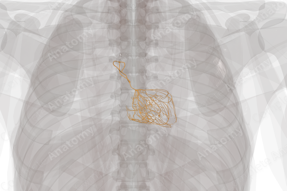
The cardiac conduction system initiates and coordinates the contraction of the myocardial tissue of the heart. This system, which receives autonomic input from the cardiac plexus, consists of highly specialized cardiac muscle cells. These low contractile, highly depolarizing cells create a unidirectional conduction pathway that travels through the walls of the heart.
The major components of the cardiac conduction system include the sinuatrial (SA) node, atrioventricular (AV) node, AV bundle, various branches and tracts that spread from the nodes throughout the heart, and Purkinje fibers that pass the action potential to the myocardial muscle.
Typically, the signal for the normal cardiac cycle to occur is initiated in the SA node, which is often referred to as the pacemaker. In the absence of autonomic input, the SA node has an inherent depolarizing cycle of roughly 70–75 beats per minute and can thus maintain heart function independently.
From the SA node, the signal spreads to atrial tissue leading to atrial contraction as well as through internodal tracts to the AV node. The AV node also has an inherent cycle of depolarization of 40–50 beats per minute. Typically, the basal rhythm of the AV node is masked by the higher pace of the SA node, but in pathologic situations, the AV node can take over pacemaker function for the heart.
Electrical signals travel from the AV node thorough the AV bundle and down the walls of the interventricular septum. Branches of this bundle distribute impulses to the Purkinje cells that are found in the innermost layer of the myocardium. Purkinje cells propagate an action potential more quickly than the rest of the cardiac conduction system. They directly trigger the process of myocardial contraction and spread the wave through from endocardial surface to epicardial surface of the heart. The electrical depolarization and repolarization generated and coordinated by the cardiac conduction system is what can be seen on an ECG in clinical practice (Anderson et al., 2009; Katz, 2010).


Related parts of the anatomy
References
Anderson, R. H., Yanni, J., Boyett, M. R., Chandler, N. J. and Dobrzynski, H. (2009) 'The anatomy of the cardiac conduction system', Clin Anat, 22(1), pp. 99-113.
Katz, A. M. (2010) Physiology of the Heart. M - Medicine Series: Wolters Kluwer Health/Lippincott Williams & Wilkins Health.
Learn more about this topic from other Elsevier products

