Quick Facts
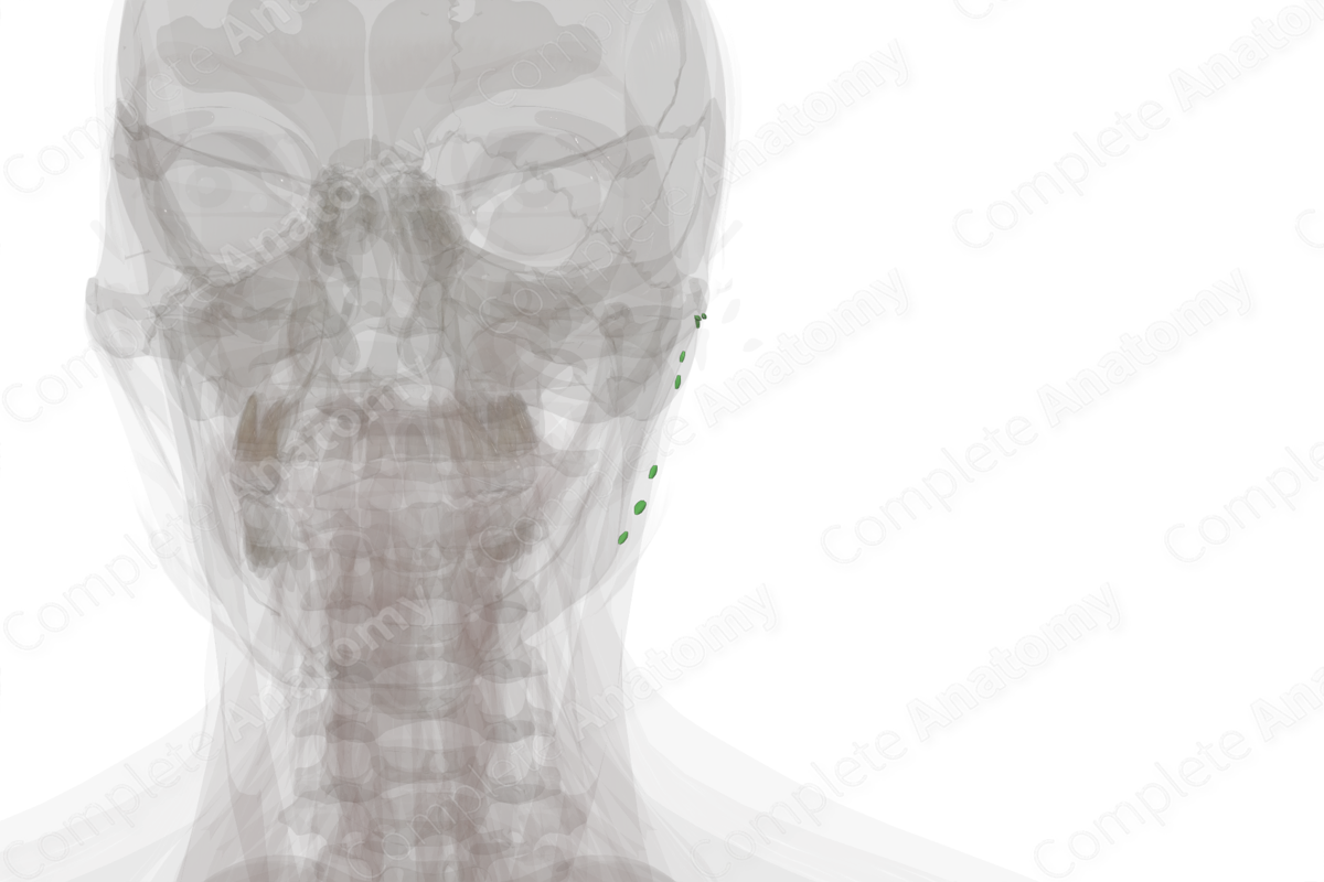
Location: On the surface of the parotid gland, with close relations to the surrounding fascia.
Drainage: The parotid gland, the frontal and temporal scalp, the nares, gingiva and the upper lip, the eyelids and external ear.
Direction of Flow: Internal jugular nodes > supraclavicular nodes > jugular trunk > thoracic duct (left) or right lymphatic duct.
Related parts of the anatomy
Description
The superficial parotid lymph nodes can be further subcategorized into preauricular and infraauricular nodes, depending on their location.
The preauricular nodes are usually one to four nodes located outside the parotid fascia (epifascial) and one to four nodes enclosed by the parotid fascia (subfascial) and adjacent to the superficial temporal artery and vein. The preauricular nodes specifically drain the superficial areas of the face and temporal region.
The infraauricular nodes are one to three nodes located inferior to the parotid gland and anteroinferior to the auricle that are located beneath the fascia of the inferior parotid pole (subfascial). They have a close anatomical relation with the retromandibular vein and can extend as far posteriorly as the tragus. In newborns, these nodes extend behind the auricle along with the parotid gland.
Drainage of both of these types of nodes is via the deep cervical nodes and jugular lymph vessels in the lateral aspect of the neck. Superficial parotid nodes are variable in number and are often too small to see without the use of a microscope (Földi et al., 2012).
List of Clinical Correlates
—Cutaneous malignancy of the ear, cheek, temple, forehead and anterior scalp
References
Földi, M., Földi, E., Strößenreuther, R. and Kubik, S. (2012) Földi's Textbook of Lymphology: for Physicians and Lymphedema Therapists. Elsevier Health Sciences.
Learn more about this topic from other Elsevier products


