Quick Facts
Location: Abdominal cavity.
Arterial Supply: Jejunal arteries, which are branches of the superior mesenteric artery.
Venous Drainage: Jejunal veins, draining to the superior mesenteric vein.
Innervation: Parasympathetic: vagus nerve (CN X); Sympathetic: celiac ganglia; Visceral afferents: spinal ganglia of T5-T9; Enteric innervation.
Lymphatic Drainage: Juxtaintestinal nodes to the central superior mesenteric nodes.
Structure/Morphology
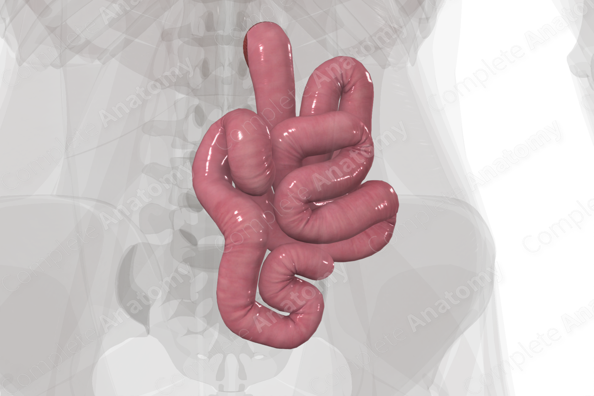
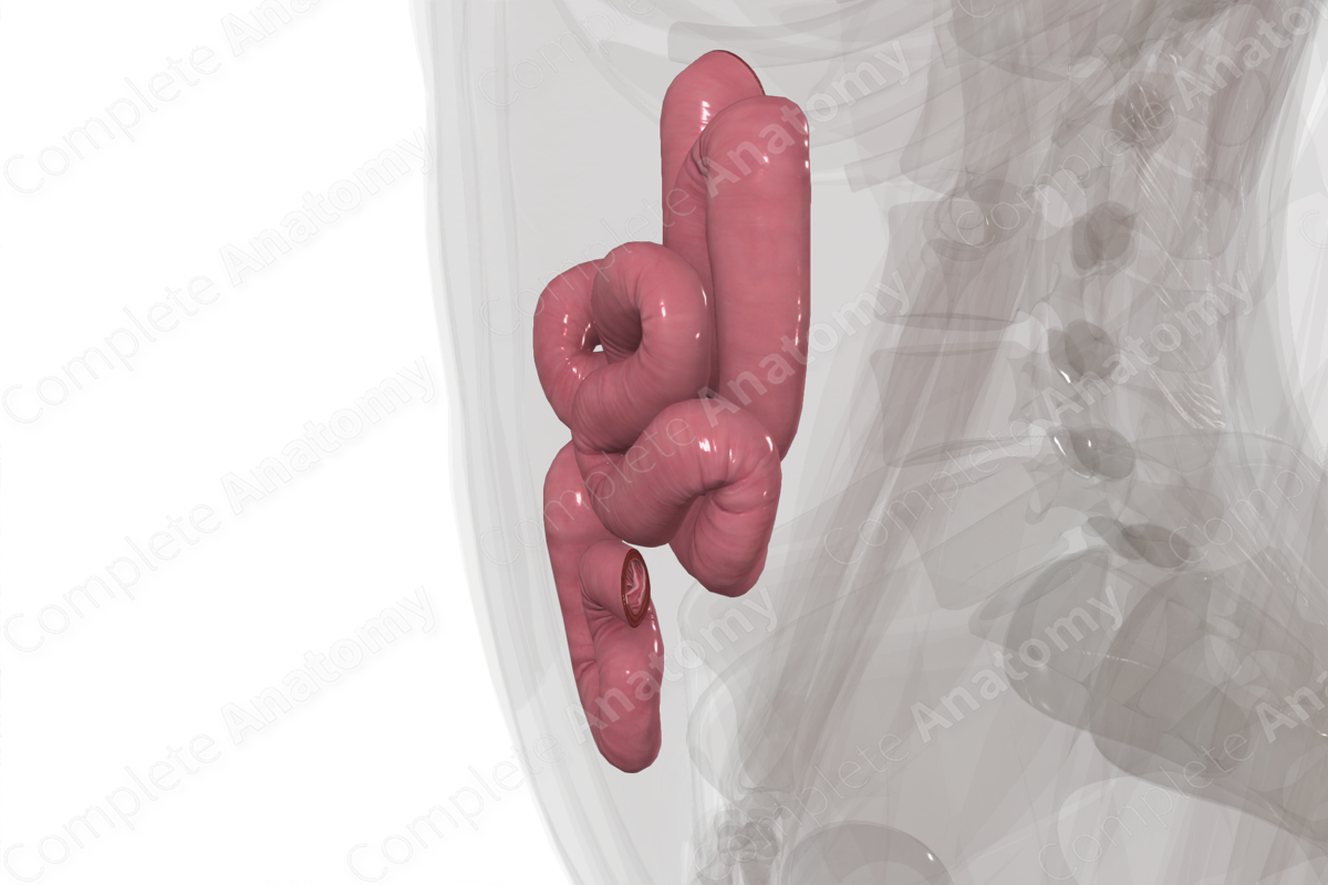
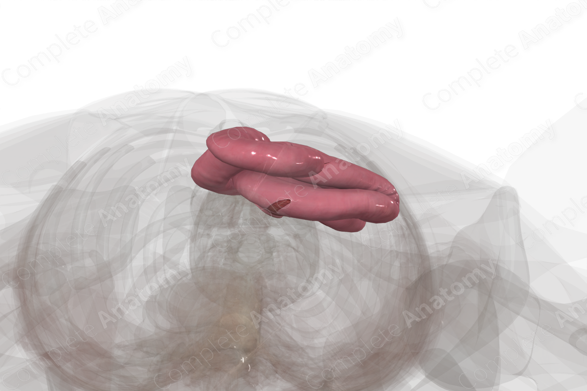
The jejunum is the second part of the small intestine, and entirely midgut. It begins at the duodenojejunal junction and ends at the ileum.
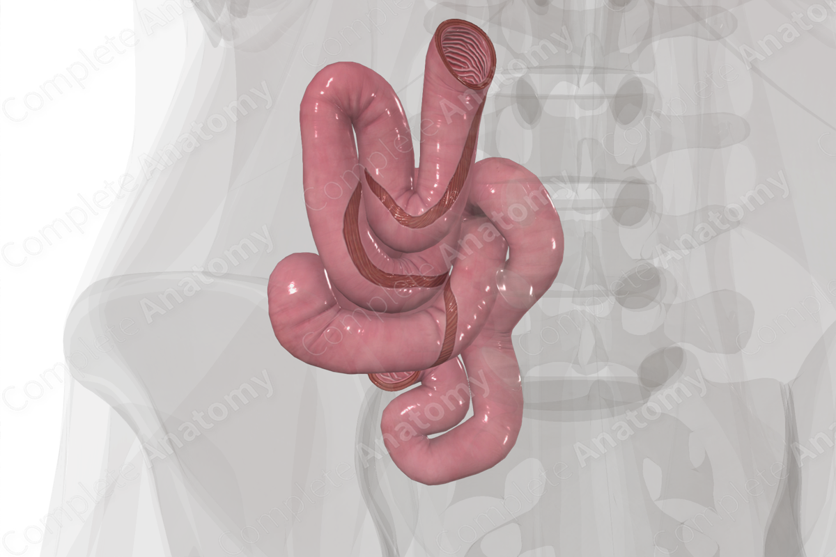
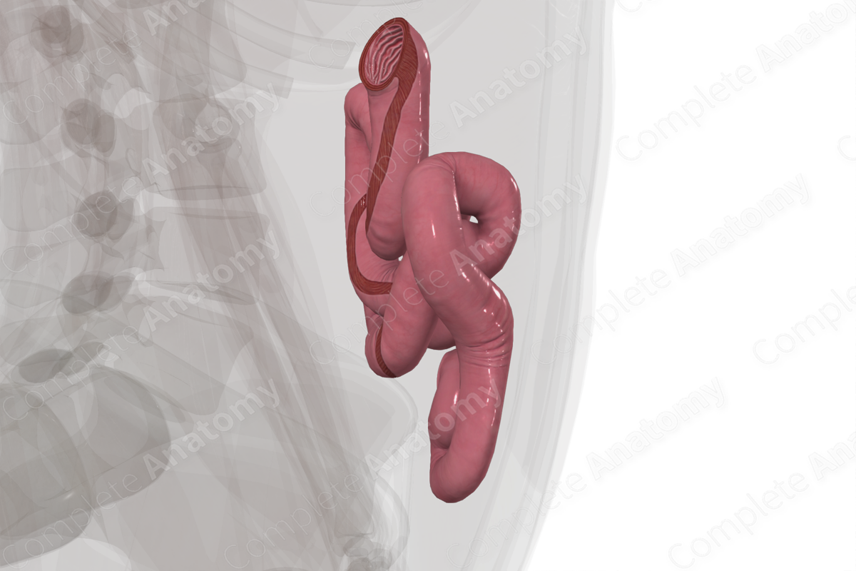
The cross-sectional microarchitecture of the jejunum demonstrates mucosa, submucosa, and muscular (inner circular and outer longitudinal) layers with a thin outer serosal covering (Standring, 2016). The jejunum has larger circular folds, villi, and a thicker mucosa than the ileum. It also has few to no Brunner’s glands (duodenum) and Peyer’s patches (ileum).
Anatomical Relations
The jejunum is the second part of the small intestine. It is intraperitoneal and sits in the abdominal cavity, deep to the umbilical region and separated from the anterior abdominal wall by the greater omentum which overlays it. The jejunum is attached to the posterior abdominal wall by the mesentery that carries the superior mesenteric vasculature.
The jejunum begins at the duodenojejunal junction just distal to the suspensory ligament of the duodenum (or ligament of Treitz), roughly ventral and left of the L2 vertebra. The jejunum is roughly 2.5-3 m long and coils within the abdominal cavity to the start of the ileum. The transition from jejunum to ileum is not well defined.
Function
The jejunum is primarily a site of absorption but final enzymatic digestion of nutrients on the brush border (microvilli) of enterocytes also occurs.
Microvilli on the enterocytes that line the lumen of the jejunum are the site of absorption of nutrients. Lipids, which are primarily absorbed in the jejunum, are shuttled into the lymph system. All other nutrients are sent into the portal venous system (Standring, 2016).
Arterial Supply
The jejunum is supplied by jejunal branches of the superior mesenteric artery.
Within the mesentery, these arteries merge to form loops that are called arcades. Arcades give rise to branches called the vasa recta that run to the jejunum itself. The arcades of the jejunum are fewer and larger than those of the ileum, while the vasa recta of the jejunum are longer than that of the ileum.
Venous Drainage
The venous drainage of the jejunum is to the portal circulation. The pattern largely parallels the arterial supply.
Nutrient rich blood draining from the jejunum drains back through jejunal veins, into the superior mesenteric vein, and into the portal vein and liver.
Innervation
Innervation of the jejunum includes the enteric nervous system (sensory and motor), the autonomic nervous system (sympathetic and parasympathetic), and extrinsic sensory innervation (visceral afferents) (Standring, 2016).
The vagus nerve (CN X) provides parasympathetic innervation to the jejunum.
Sympathetic innervation originates in the T5-T9 spinal cord levels with preganglionic neurons. The jejunum is supplied by postganglionic neurons from the celiac plexus and superior mesenteric plexus.
Visceral afferent nerves from the jejunum ascend along the vagus nerve or greater splanchnic nerve.
The enteric system consists of two plexuses of densely packed small neurons. Meissner’s plexus lies in the submucosal layer and Auerbach’s myenteric plexus lie between the outer longitudinal and inner circular smooth muscle layers. These systems of nerves control mucosal and peristaltic function.
Lymphatic Drainage
Lymphatic drainage from the jejunum parallels the arterial supply and drains from juxtaintestinal nodes into the central superior mesenteric lymph nodes. (Földi et al., 2012).
List of Clinical Correlates
- Crohn’s disease
- Jejunal diverticulosis
- Celiac Disease
- Jejunal atresia
References
Földi, M., Földi, E., Strößenreuther, R. and Kubik, S. (2012) Földi's Textbook of Lymphology: for Physicians and Lymphedema Therapists. Elsevier Health Sciences.
Standring, S. (2016) Gray's Anatomy: The Anatomical Basis of Clinical Practice. Gray's Anatomy Series 41 edn.: Elsevier Limited.
Learn more about this topic from other Elsevier products

