Quick Facts
Location: Abdominal cavity.
Arterial Supply: Cystic artery, branch of right hepatic artery.
Venous Drainage: Cystic veins to portal vein; small veins to segmental portal veins in the liver.
Innervation: Parasympathetic: Vagus nerve (CN X); Sympathetic: celiac plexus; Visceral afferents: spinal ganglia of T7-T9.
Lymphatic Drainage: Cystic node and node of anterior border of omental foramen.
Related parts of the anatomy
Structure/Morphology
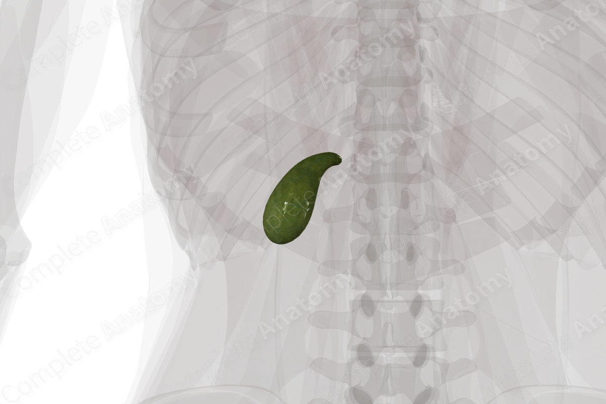
The gallbladder is a pear-shaped, blind-end diverticulum that stores and concentrates up to 50 ml of bile. It consists of three parts:
-the fundus is the rounded end;
- the body is the main part;
- the neck which connects the rest of the gallbladder to the cystic duct.
The gallbladder has layers like that of the digestive canal.
A single layer of epithelium lines the lumen and is part of the mucosal layer. The mucosa has small rugae that flattens as the gallbladder fills and expands. In the neck, the mucosa forms into spiral folds which are also found in the cystic duct and are thought to help keep the opening to the duct patent.
A muscular layer sits deep to the mucosal layer. Here smooth muscle fibers in longitudinal, circular, and oblique orientations are intermixed with connective tissue.
The muscular layer is wrapped in an adventitia composed of loose connective tissue, adipose tissue, and neurovasculature. Overlying this is the serosal layer. The serosa of the gallbladder covers most of the gallbladder, except for the intrahepatic portion (Standring, 2016).
Anatomical Relations
The gallbladder is a pear-shaped diverticulum connected by the cystic duct to the hepatic and bile ducts. It sits in a depression in the inferior surface of the liver, roughly between the right lobe and quadrate lobe. It’s often so tightly adhered to the liver that no visceral peritoneum is found between them.
The fundus often protrudes from underneath the anterior edge of the right lobe of the liver, just medial to the costal cartilage of the ninth rib. The body and fundus are often found anterior to the first parts of the duodenum.
The neck of the gallbladder narrows to become the short cystic duct.
Function
The gallbladder serves to store and concentrate bile that is produced in the liver. When the bile duct is closed, bile produced in the liver is shunted through the cystic duct for storage. The epithelial lining absorbs water to concentrate the bile. When needed, bile is sent from the gallbladder back through the cystic duct to the bile duct and duodenum to participate in digestion.
Arterial Supply
The gallbladder is supplied with blood from the cystic artery, a branch most often of the right hepatic artery.
Near the neck of the gallbladder, the cystic artery typically splits into a deep branch that runs along the superior or hepatic surface, and a superficial branch that runs down the inferior or duodenal surface of the gallbladder. The territories of these arteries spread out and anastomose with each other.
Venous Drainage
The gallbladder isn’t drained by a single vein. Instead, the bulk of the gallbladder that lies against the liver is drained through many small unnamed veins which send blood into segmental portal veins and sinusoids found within the liver (Moore, Dalley and Agur, 2013).
The “free” surfaces of the gallbladder are typically drained by several cystic veins. These send blood to the veins associated with the biliary ducts, and from there into the portal vein or its tributaries (Standring, 2016).
Innervation
The gallbladder is innervated by parasympathetic and sympathetic efferent fibers and visceral afferent sensory fibers.
The vagus nerve, particularly the anterior trunk, send parasympathetic fibers to the gallbladder via the hepatic plexus. These fibers together with hormonal signals from the duodenum cause contraction of gallbladder smooth muscle fibers.
Sympathetic efferent fibers from the greater and lesser splanchnic nerve synapse in the celiac and hepatic ganglia before sending postganglionic sympathetic fibers to the gallbladder (Standring, 2016). Their function is likely related to vasoconstriction.
Visceral sympathetic fibers from the gallbladder carry pain sensation back along the vagus nerve or the splanchnic nerves. This can be interpreted as referred pain that is often felt in the right hypochondrium and epigastrium (Standring, 2016).
Lymphatic Drainage
Lymph from the gallbladder can drain to the celiac nodes, posterior pancreaticoduodenal nodes, or the livers lymphatic ducts.
Much of the body and fundus will drain to the cystic node, a node found near the neck of the gallbladder, or the node of the anterior border of the omental foramen. From here, lymph drains through the posterior pancreaticoduodenal lymph nodes and to the celiac nodes (Földi et al., 2012).
Lymph from tissue next to the liver can drain directly into the liver and the lymphatic ducts of the liver (Standring, 2016).
List of Clinical Correlates
- Gallstones
- Cholecystitis
References
Földi, M., Földi, E., Strößenreuther, R. and Kubik, S. (2012) Földi's Textbook of Lymphology: for Physicians and Lymphedema Therapists. Elsevier Health Sciences.
Standring, S. (2016) Gray's Anatomy: The Anatomical Basis of Clinical Practice. Gray's Anatomy Series 41 edn.: Elsevier Limited.
Learn more about this topic from other Elsevier products
Gallbladder histology: Video, Causes, & Meaning

Gallbladder histology: Symptoms, Causes, Videos & Quizzes | Learn Fast for Better Retention!


