Structure/Morphology
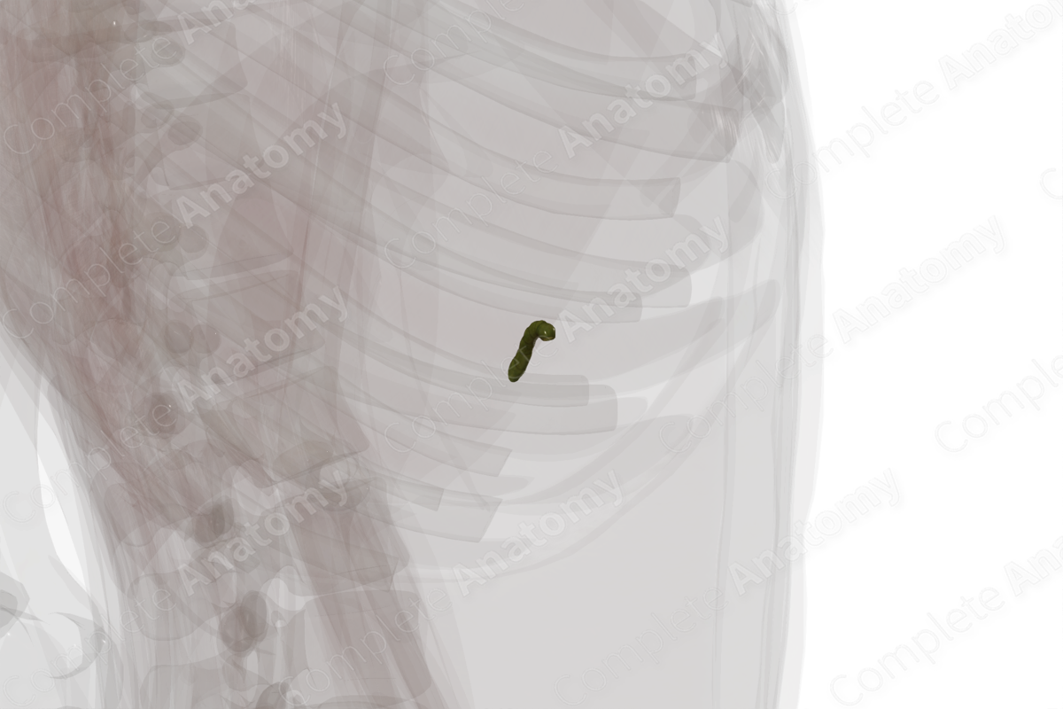
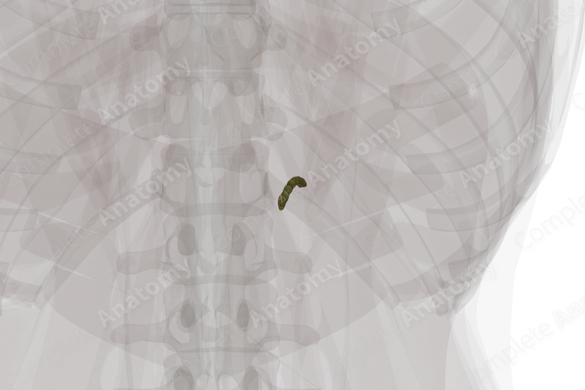
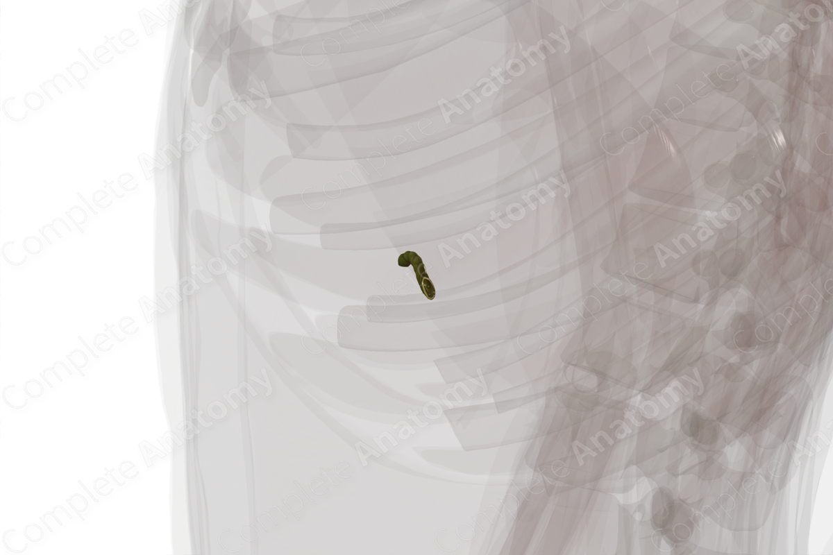
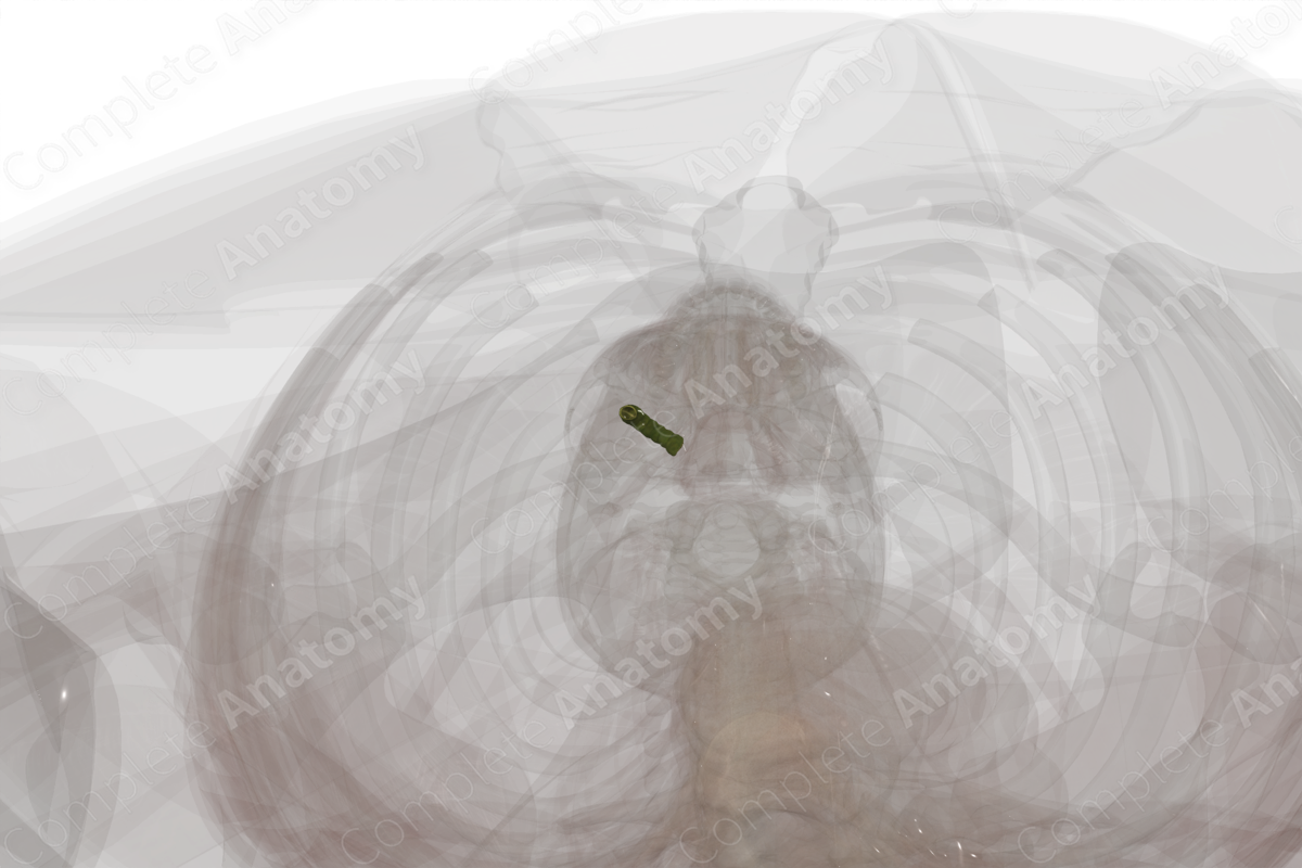
The cystic duct is a short often tortuous duct that connects the gallbladder to the bile duct and common hepatic duct.
Internally, the cystic duct has a mucosal layer containing mucus glands and an epithelium.
Externally, the cystic duct has a fibrous layer containing fibrous connective tissue and smooth muscle cells found in several orientations (Standring, 2016).
Related parts of the anatomy
Key Features/Anatomical Relations
The cystic duct runs 2–4 cm from the neck of the gallbladder to the junction with the hepatic duct. Thus, the duct running towards the liver is the common hepatic duct, while the duct distal to this junction is the bile duct.
A prominent feature is the specialized mucosal folds called spiral folds or the spiral valves of Heister. These folds, which are also found in the neck of the gallbladder, are thought to help keep the cystic duct open to allow for passage of bile.
Function
The cystic duct connects the gallbladder to the biliary system. It allows bile from the liver to pass into the gallbladder for storage and concentration and allows bile from the gallbladder to be sent to the duodenum in response to hormonal signals during digestion.
References
Standring, S. (2016) Gray's Anatomy: The Anatomical Basis of Clinical Practice. Gray's Anatomy Series 41 edn.: Elsevier Limited.
Learn more about this topic from other Elsevier products


