Description
The retroperitoneal space is the abdominal compartment resting between the parietal peritoneum anteriorly and the fascia covering the musculature of the posterior abdominal wall.
The diaphragm marks the superior margin of the retroperitoneal space and the extraperitoneal fascia, lining the muscles that form the pelvic floor, mark the inferior margin. Laterally, the retroperitoneal space ends where the parietal peritoneum reflects anteriorly along the body wall. Here it aligns with the transversalis fascia lining the body wall musculature, leaving only potential space between them (Standring, 2016).
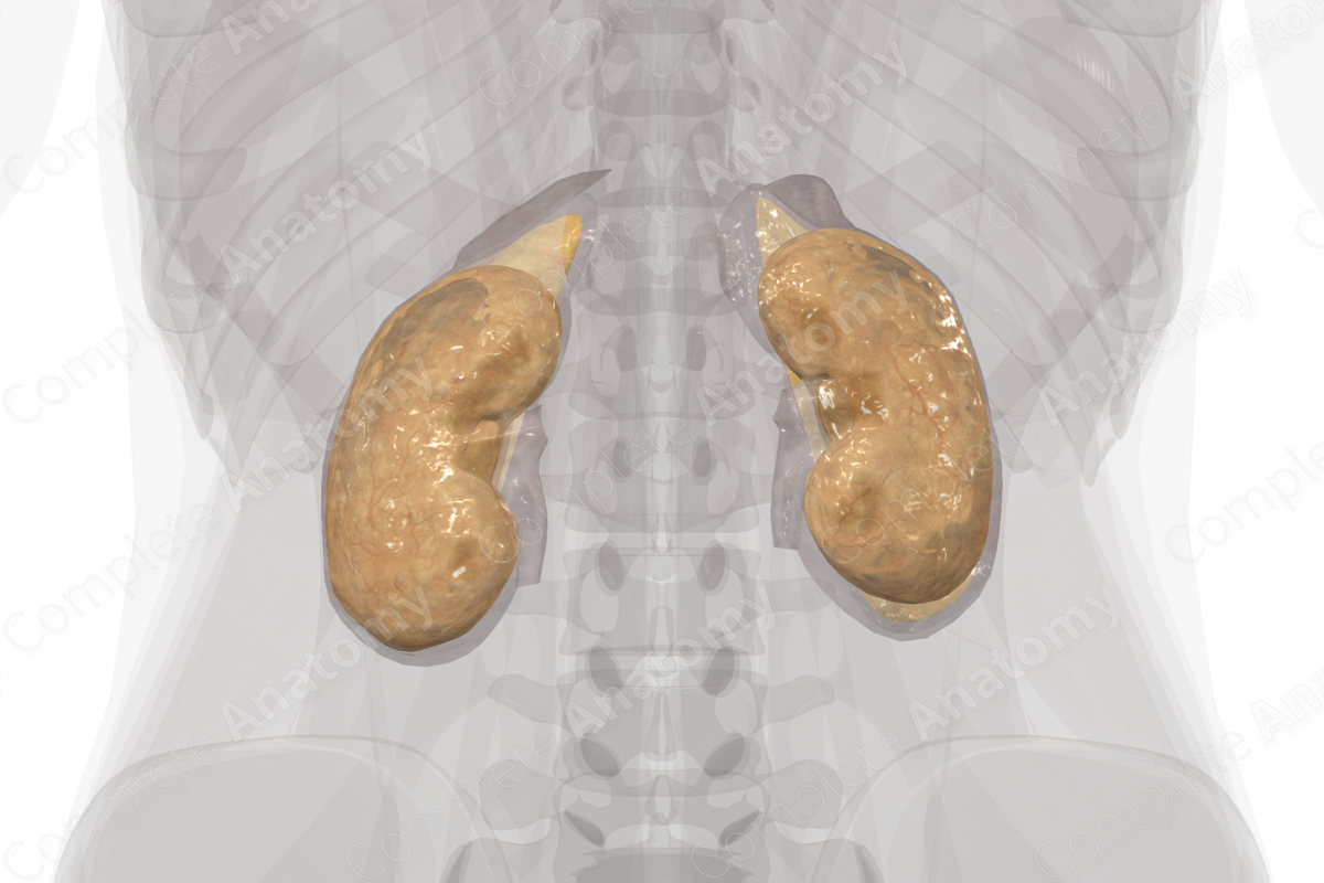
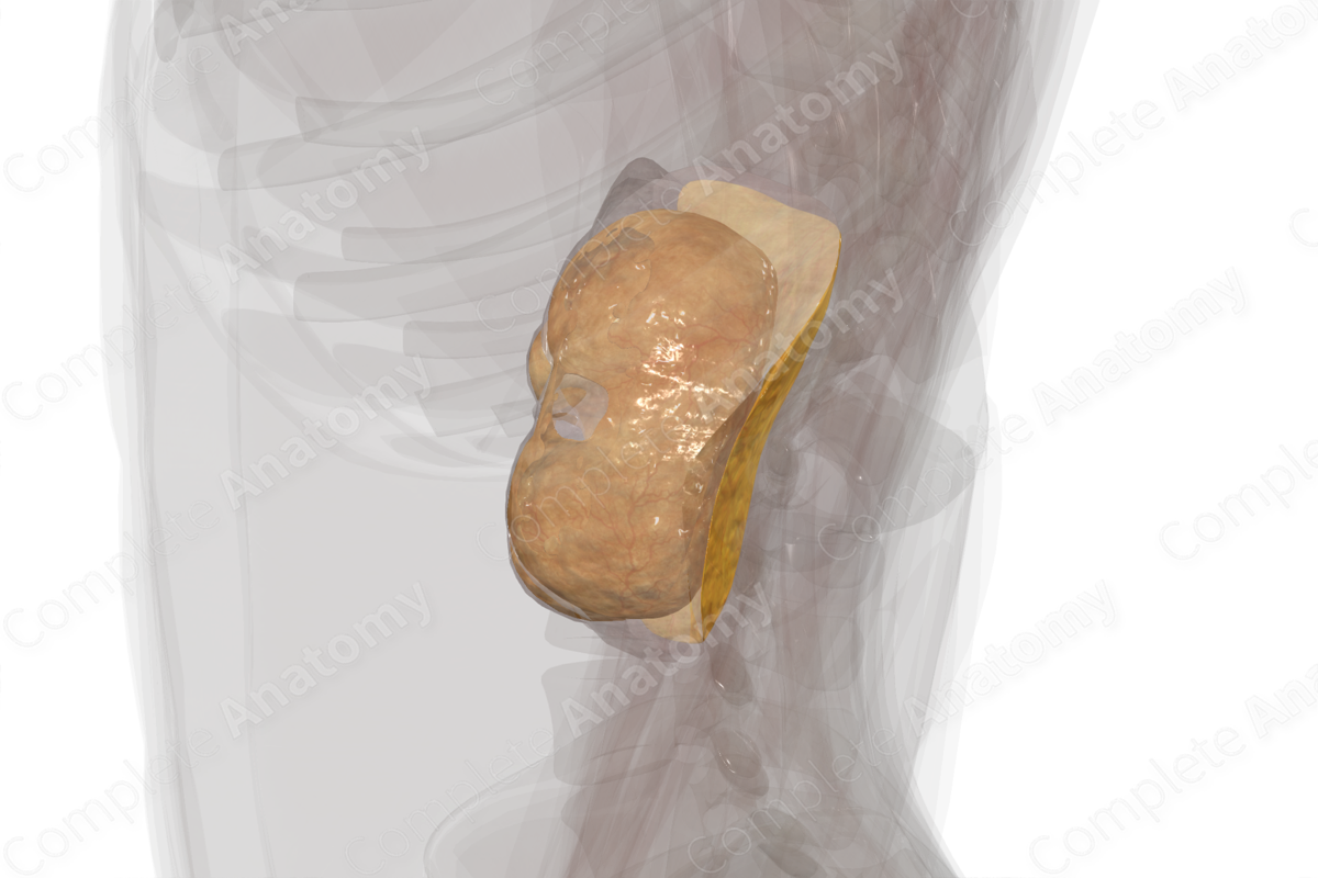
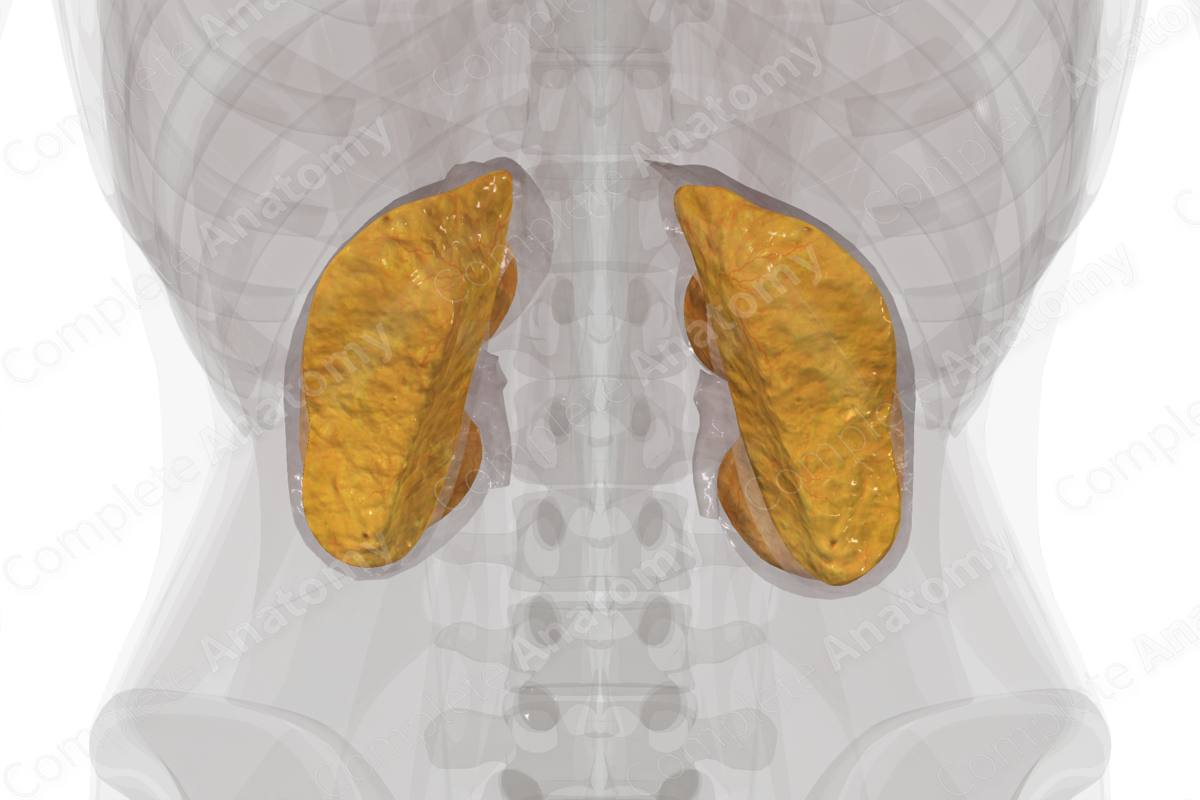
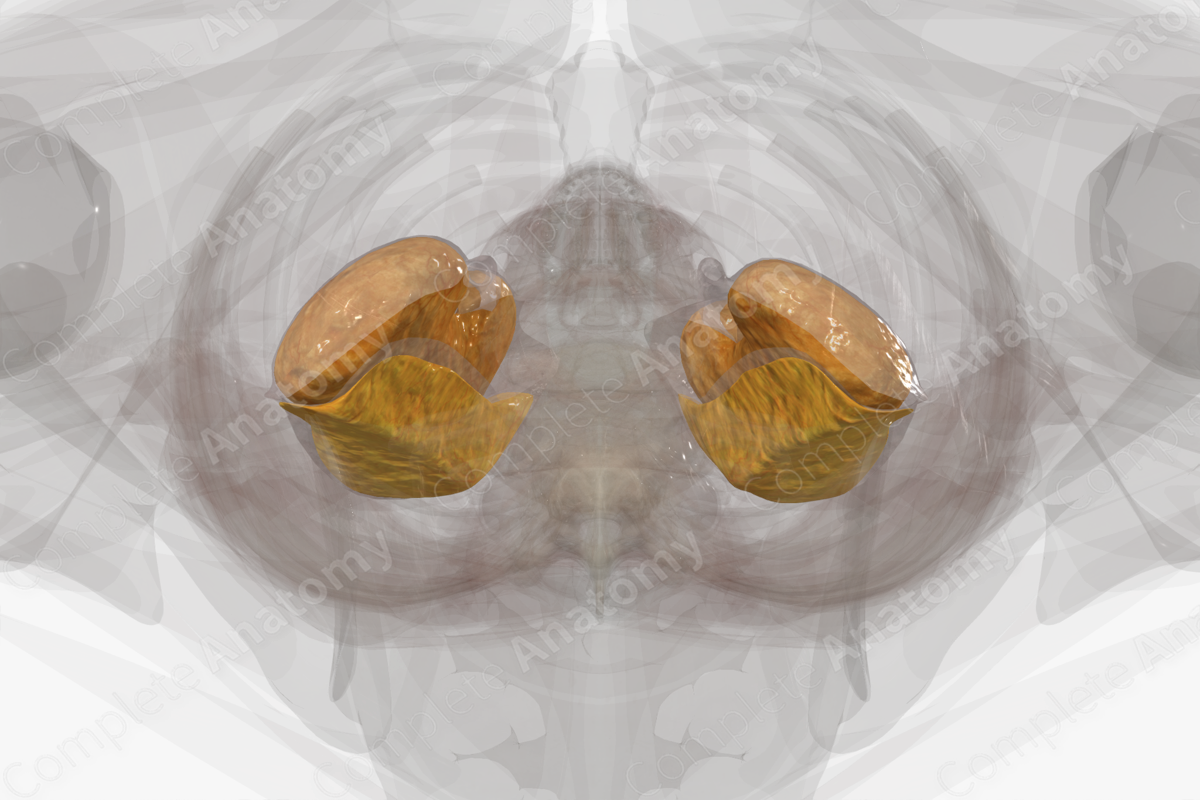
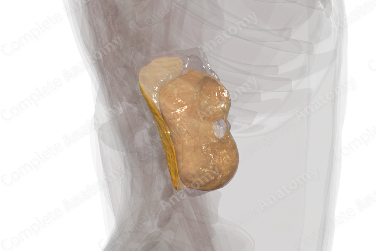
Found within the retroperitoneal space are the nerves, arteries, veins, and lymphatics serving the abdominal wall and viscera of both the abdominal and pelvic cavities, as well as retroperitoneal space. The kidneys, adrenal glands, and ureters form within the retroperitoneal space, enclosed by a renal fascia. Secondarily retroperitoneal organs, such as portions of the large intestine and duodenum, as well as the pancreas, migrate during development from an intraperitoneal origin back into the retroperitoneal space. Running within the retroperitoneal space are the abdominal aorta and the inferior vena cava, and branches of both vessels that serve organs and tissues in this space. Spinal nerves of the lumbosacral plexus and autonomic nerves serving the viscera of the abdomen also run in the retroperitoneal space (Mirilas and Skandalakis, 2009).
Those nerves and vessels that serve intraperitoneal organs run in the retroperitoneal space until, at the appropriate location, they exit the retroperitoneum via a mesentery to reach their intraperitoneal target.
References
Mirilas, P. and Skandalakis, J. E. (2009) 'Surgical anatomy of the retroperitoneal spaces--part I: embryogenesis and anatomy', Am Surg, 75(11), pp. 1091-7.
Standring, S. (2016) Gray's Anatomy: The Anatomical Basis of Clinical Practice. Gray's Anatomy Series 41st edn.: Elsevier Limited.
Learn more about this topic from other Elsevier products

