Quick Facts
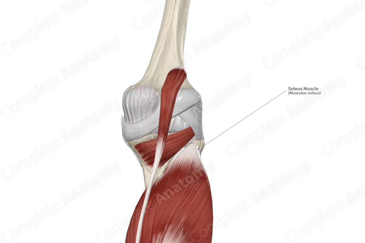
Origin: Head of fibula, posterior surface of fibula, soleal line, and medial border of tibia.
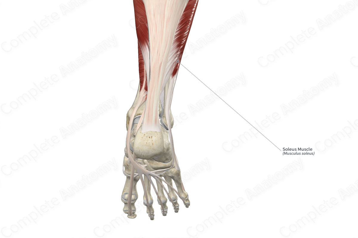
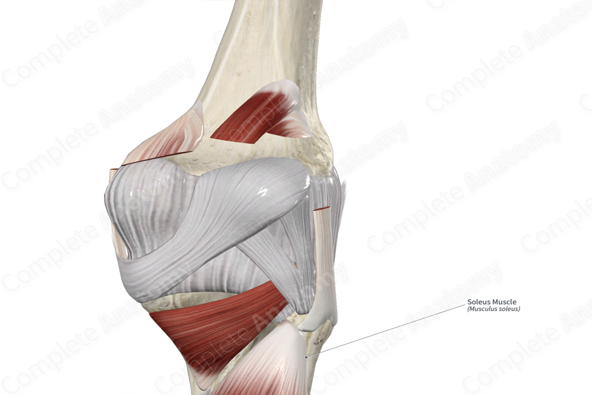
Insertion: Posterior surface of calcaneus, via calcaneal tendon.
Action: Plantarflexes foot at ankle joint.
Innervation: Tibial nerve (S1-S2).
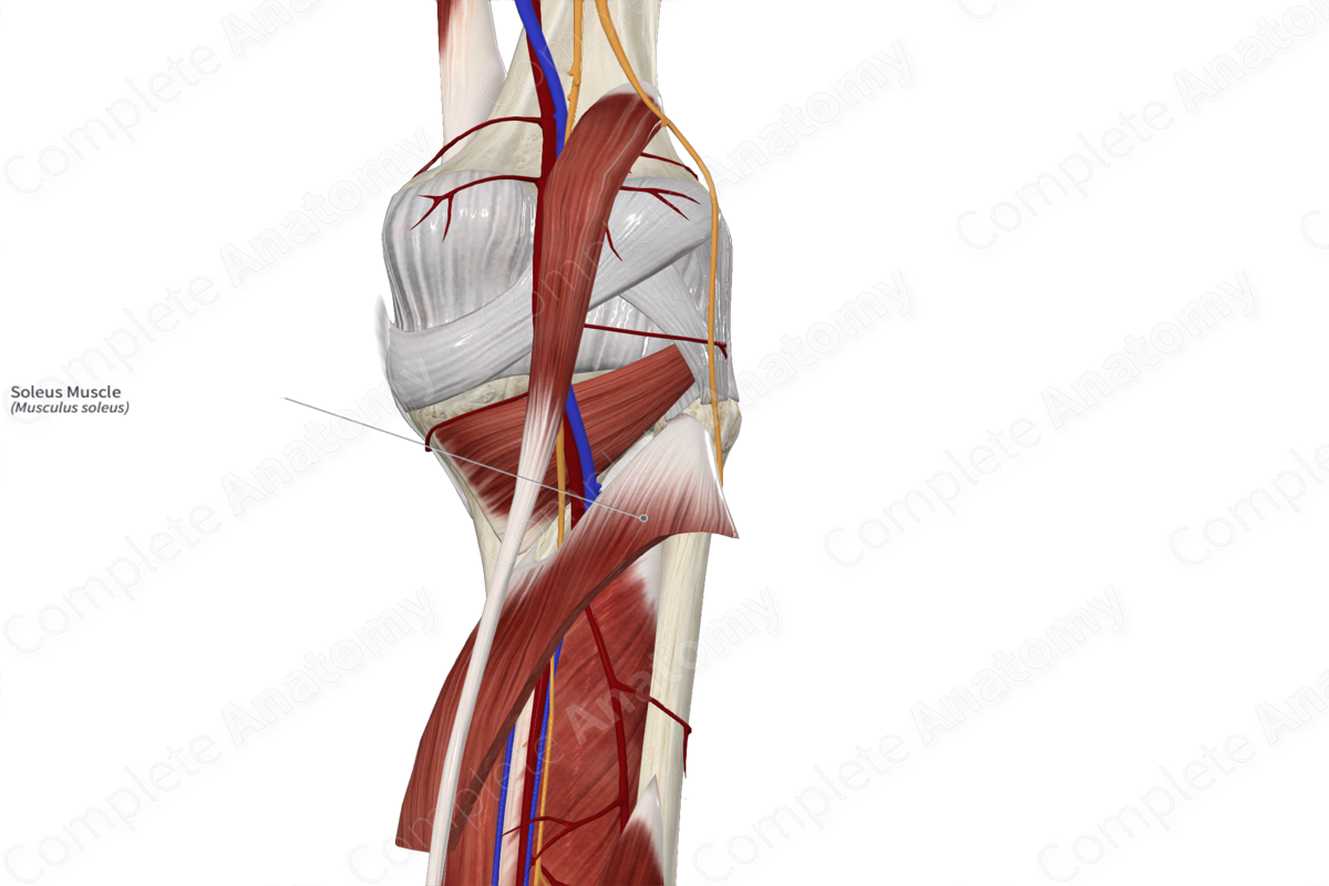
Arterial Supply: Popliteal, fibular, and posterior tibial arteries.
Related parts of the anatomy
Origin
The soleus muscle originates from the:
- posterior aspect of the head and neck of fibula;
- proximal one quarter of the posterior surface of fibula;
- soleal line of tibia;
- middle one third of the medial border of tibia.
The soleus muscle also originates from a fibrous band that extends from its origin sites on the fibula and tibia, known as the tendinous arch of soleus.
Insertion
The fibers of the soleus muscle travel inferiorly and converge with the fibers of the medial and lateral heads of the gastrocnemius muscle to form the calcaneal tendon, which inserts onto the posterior surface of the calcaneus.
Key Features & Anatomical Relations
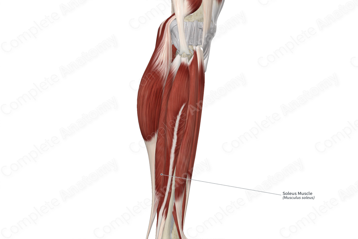
The soleus muscle is one of the three muscles that form the triceps surae muscle, the other two being the medial and lateral heads of the gastrocnemius muscle. It is a broad, flat, multipennate type of skeletal muscle.
It is located:
- anterior (deep) to the medial and lateral heads of the gastrocnemius muscle and the tendon of the plantaris muscle;
- posterior (superficial) to the transverse intermuscular septum of leg, the flexor digitorum longus, flexor hallucis longus and tibialis posterior muscles, the posterior tibial vessels, and the tibial nerve;
- lateral to the great saphenous vein and the saphenous nerve.
In order to enter the posterior compartment of the leg, the popliteal vessels and tibial nerve travel deep to the tendinous arch of soleus muscle.
Actions & Testing
The soleus muscle is involved in multiple actions:
- plantarflexes the foot at the ankle joint;
- helps stabilize the leg on the foot during standing.
The soleus muscle cannot be tested in isolation, therefore all three muscles of the triceps surae are tested simultaneously by plantarflexing the foot at the ankle joint against resistance, during which both the soleus muscle and calcaneal tendon can be palpated (Sinnatamby, 2011).
List of Clinical Correlates
- Deep vein thrombosis
References
Sinnatamby, C. S. (2011) Last's Anatomy: Regional and Applied. ClinicalKey 2012: Churchill Livingstone/Elsevier.
Learn more about this topic from other Elsevier products

